
MCU/ IVP / AUG
MCU/ IVP / AUG
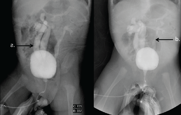
The difference between the cystogram and a urethrogram is that towards the end of the procedure, once the catheter is removed, you will be asked to pass urine into a pan or bottle. The Radiologist will watch on the x-ray screen to see how the size and shape of the bladder changes during urination and image the urethra also. The upper urinary tract (Ureter and kidneys) are imaged to assess for any reflux of contrast back up the ureters into the kidneys.
No Catheter in place
- The catheter is a small tube which the Radiologist uses to introduce a mixture of contrast media and saline into the bladder.
- The Radiologist will first cleanse the urethral area with Savlon, a numbing gel will be used and then pass a small tube through the urethra into the bladder. This may sting a little.
- The contrast media is then introduced and the Radiologist will watch on the x-ray screen as the bladder fills. More images may be taken in different positions.
- The catheter will now be removed. You will be sent to the rest room to empty your bladder.
Catheter already in place
- If you have a catheter in place, the Radiologist will use this to administer the contrast media. The contrast media is introduced and the Radiologist will watch on the x-ray screen as the bladder fills. More images may be taken in different positions.
- On completion of the examination they catheter may be removed or may stay in place. The referring specialist will communicate with the Radiologist about this. The outcome is often dependent on the result. The contrast may be removed either by syringe or by draining into your catheter bag.