
Understanding Fatty Liver Detection Through Ultrasound and MRI
Fatty liver disease has quietly become one of the most common health concerns in India, often discovered almost by accident — during a routine health check-up, an unrelated abdominal scan, or a doctor’s visit for vague symptoms like fatigue or mild discomfort. The tricky part about fatty liver is that it rarely announces itself early on. By the time symptoms appear, the condition may have already progressed.
This is exactly why a timely fatty liver scan matters. Imaging technology — particularly ultrasound and MRI — plays a central role in catching fatty liver early, tracking its progression, and helping doctors decide the right course of action. At Sunray Scans, Chennai, patients have access to both modalities under one roof, backed by expert radiologists and fast, accurate reporting.
In this blog, we’ll walk through what fatty liver disease actually is, how liver ultrasound and liver MRI work for detection, what each scan can and can’t tell you, and why choosing the right diagnostic center matters just as much as choosing the right scan.
What Is Fatty Liver Disease?
Fatty liver disease occurs when excess fat builds up in liver cells. A small amount of fat in the liver is normal, but when fat accounts for more than 5–10% of the liver’s weight, it’s classified as fatty liver, medically known as hepatic steatosis.
There are two broad categories:
- Non-Alcoholic Fatty Liver Disease (NAFLD) — linked to obesity, diabetes, high cholesterol, and sedentary lifestyles, and increasingly common even in people who don’t drink alcohol at all
- Alcoholic Fatty Liver Disease (AFLD) — caused by excessive alcohol consumption over time
NAFLD, in particular, has seen a sharp rise due to changing diets, increased screen time, and reduced physical activity — making liver health screening far more relevant today than it was even a decade ago.
Left unmanaged, fatty liver can progress through stages: simple fatty liver → non-alcoholic steatohepatitis (NASH) with inflammation → fibrosis → cirrhosis. The earlier it’s caught, the more reversible it tends to be — which is precisely where imaging comes in.
Why Early Fatty Liver Diagnosis Matters
Most people with fatty liver disease don’t experience obvious symptoms in the early stages. Some may notice:
- Mild fatigue
- A dull ache or discomfort in the upper right abdomen
- General weakness
- Unexplained weight changes
Because these signs are so non-specific, fatty liver is often picked up incidentally during imaging done for other reasons. This makes fatty liver diagnosis through imaging not just a confirmatory tool, but often the first real signal that something needs attention — long before blood tests or symptoms would suggest a problem.
Liver Ultrasound: The First Line of Detection
For most patients, liver ultrasound is the starting point for fatty liver evaluation, and for good reason. It’s non-invasive, doesn’t involve radiation, is widely available, and provides reasonably reliable results for moderate to severe fatty liver.
How Ultrasound Detects Fatty Liver
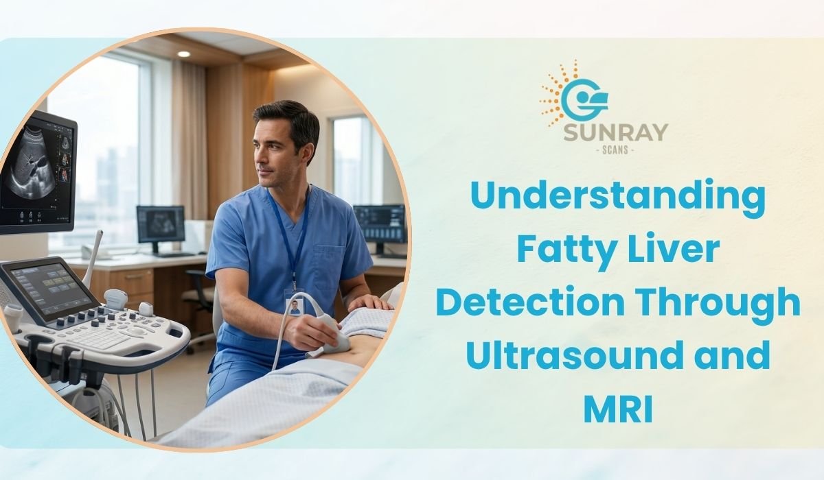
During an abdominal ultrasound, sound waves are used to create real-time images of the liver. A healthy liver typically appears with a uniform, moderate echo pattern. When fat accumulates in liver tissue, it changes how sound waves reflect back, producing a characteristic “bright” or hyperechoic appearance on the screen.
Radiologists also look for:
- Increased liver size (hepatomegaly)
- Reduced visibility of liver blood vessels due to fat interference
- Difference in brightness between the liver and the right kidney, a classic comparative marker
- Attenuation of the ultrasound beam as it passes deeper into liver tissue
Grading Fatty Liver on Ultrasound
Ultrasound typically grades fatty liver into three categories:
- Grade I (Mild): Slightly increased liver brightness, vessels still visible
- Grade II (Moderate): More noticeable brightness, partial loss of vessel and diaphragm visibility
- Grade III (Severe): Significant brightness, poor visualization of deeper liver structures and diaphragm
Strengths and Limitations of Ultrasound for Fatty Liver
Ultrasound is excellent as a first-line, accessible screening tool, but it does have limitations:
- It’s less sensitive for detecting mild fat infiltration (below roughly 20%)
- Results can be operator-dependent, which is why experienced radiologists matter significantly
- It doesn’t precisely quantify the exact percentage of fat in liver tissue
- Accuracy can be affected by patient body type, particularly in cases of obesity
This is why, for more detailed evaluation or when ultrasound findings are inconclusive, doctors often recommend stepping up to MRI-based abdominal imaging.
Liver MRI: The More Detailed Diagnostic Step
When precision matters — whether for early-stage detection, monitoring disease progression, or differentiating fatty liver from other liver conditions — liver MRI offers a level of detail ultrasound simply can’t match.
How MRI Detects and Quantifies Fatty Liver
MRI uses magnetic fields and radio waves to generate highly detailed cross-sectional images of the liver without any radiation exposure. For fatty liver specifically, a specialized technique called MRI-PDFF (Proton Density Fat Fraction) has become something of a gold standard in modern hepatology.
What makes MRI particularly valuable:
- It can detect fat infiltration as low as 5%, far more sensitive than ultrasound
- It provides a quantifiable fat percentage, not just a qualitative grade
- It can help distinguish fatty liver from other structural liver changes
- It’s largely unaffected by patient body habitus, unlike ultrasound
- Advanced MRI sequences can also assess for early fibrosis in some cases, giving doctors a broader picture of liver health beyond just fat content
Open MRI: A More Comfortable Diagnostic Experience
One challenge with traditional MRI machines is the closed, tunnel-like design, which can cause significant discomfort or anxiety for many patients — particularly those with claustrophobia, larger body frames, or younger and elderly patients who struggle to stay still in confined spaces.
Sunray Scans addresses this directly with Chennai’s first High-Resolution Open MRI, built on HTS coil technology that delivers sharp, diagnostic-quality images without the closed, tunnel-like feel of conventional machines. The open, spacious design and quieter operation make it considerably easier for patients to stay relaxed and still throughout the scan — which, in turn, supports clearer, more reliable imaging for conditions like fatty liver.
Ultrasound vs MRI: Which One Do You Need?
Patients often ask whether they should go straight for an MRI or start with an ultrasound. In practice, it usually comes down to the clinical context:
Factor | Ultrasound | MRI |
Best for | Initial screening, moderate-severe fatty liver | Precise quantification, early-stage detection |
Sensitivity for mild fat | Lower | Higher |
Radiation | None | None |
Cost | Lower | Higher |
Speed | Faster | Takes longer |
Operator dependency | Higher | Lower |
For most routine liver health screening, ultrasound remains the practical first step — it’s quick, affordable, and effective for moderate to advanced fatty liver. MRI becomes especially valuable when:
- Ultrasound results are unclear or borderline
- A patient is being monitored for treatment response over time
- A more precise fat quantification is needed for clinical decision-making
- There’s a need to rule out other liver abnormalities alongside fatty liver
A good diagnostic center should be able to guide you toward whichever scan is clinically appropriate, rather than defaulting to the more expensive option unnecessarily.
What Happens During the Scan at Sunray Scans
For patients preparing for a fatty liver scan, the process is straightforward:
For Ultrasound:
- A short fasting period (usually 6–8 hours) is generally recommended for clearer abdominal visualization
- The scan itself typically takes 15–20 minutes
- It’s painless and involves no preparation beyond fasting
For MRI (including Open MRI):
- Patients are guided through the process by trained technicians to ensure comfort
- The open design minimizes the closed-in feeling associated with conventional MRI
- Imaging is reviewed by experienced radiologists for accurate interpretation
Sunray Scans, located in Royapettah, Chennai, combines both ultrasound and advanced Open MRI capabilities at a single facility, supported by a team of experienced radiologists and a strong tele-radiology reporting network for fast, dependable interpretation.
Beyond the Scan: What Happens After Diagnosis
Getting a fatty liver scan is just the first step. Once fat infiltration is confirmed and graded, the next steps typically involve a physician evaluating:
- Liver function tests (blood work)
- Blood sugar and cholesterol levels
- Body weight and metabolic health markers
- Alcohol consumption history
- Possible need for further evaluation like a FibroScan or liver biopsy in advanced cases
Imaging gives doctors the visual evidence; clinical correlation with blood work and patient history completes the picture. This is why a fatty liver diagnosis should always be discussed with your physician rather than self-interpreted from a scan report alone.
Lifestyle Factors That Influence Fatty Liver
While this blog focuses primarily on detection, it’s worth noting that fatty liver — especially in its early, non-alcoholic form — is often significantly influenced by lifestyle factors such as:
- Sedentary routines and lack of regular physical activity
- Diets high in refined sugars and saturated fats
- Obesity and insulin resistance
- Poorly controlled diabetes or high cholesterol
- Excessive alcohol consumption
The encouraging part is that early-stage fatty liver is often reversible with consistent lifestyle changes — another strong reason why early detection through imaging carries so much weight.
Why Choosing the Right Scan Center Matters
Not all diagnostic centers offer the same level of accuracy or patient experience. When it comes to something as nuanced as fatty liver diagnosis, a few things make a real difference:
- Equipment quality: Modern ultrasound and MRI machines produce clearer, more diagnostically useful images
- Radiologist expertise: Since ultrasound especially is operator-dependent, experienced radiologists significantly improve diagnostic accuracy
- Patient comfort: A stressed or uncomfortable patient may move during scanning, affecting image quality — this is part of why Open MRI technology has become so valuable
- Turnaround time: Faster, reliable reporting helps your doctor begin treatment planning sooner
Sunray Scans brings these elements together — combining high-resolution Open MRI, advanced ultrasound, and a team of expert radiologists, with same-day reports for most scans to support quicker diagnosis and treatment planning.
Frequently Asked Questions
Is fasting required before a fatty liver ultrasound?
Yes, most centers recommend 6–8 hours of fasting before an abdominal ultrasound. Food intake can cause gas buildup in the intestines, which interferes with clear visualization of the liver and surrounding organs.
Can fatty liver be detected on a routine abdominal ultrasound?
In many cases, yes. Moderate to severe fatty liver is often picked up incidentally during a routine abdominal ultrasound done for unrelated reasons, which is one of the most common ways the condition is first identified.
Does a liver MRI involve radiation?
No. MRI uses magnetic fields and radio waves rather than ionizing radiation, making it a safe option for repeated monitoring over time if your doctor recommends follow-up scans.
How long does an Open MRI scan take?
Duration depends on the specific protocol your doctor requests, but liver imaging typically takes longer than an ultrasound. The open design at Sunray Scans is built to make this longer duration considerably more comfortable for patients.
Can fatty liver be reversed once detected?
Early-stage fatty liver, particularly NAFLD, often responds well to sustained lifestyle changes such as improved diet, regular physical activity, and better management of blood sugar and cholesterol. This is best discussed with your treating physician based on your scan results and overall health profile.
Do I need both an ultrasound and an MRI? Not necessarily. Many patients only need an ultrasound for routine screening. An MRI is generally recommended when more precise fat quantification is needed, when ultrasound results are inconclusive, or when a doctor wants to monitor disease progression closely.
Final Thoughts
Fatty liver disease may develop silently, but it doesn’t have to stay hidden. With the right combination of liver ultrasound and liver MRI, doctors can detect fat infiltration early, track its progression accurately, and guide patients toward timely intervention — often well before the condition becomes serious.
Whether you’re going in for a routine liver health screening or following up on a doctor’s recommendation after unclear symptoms, the right abdominal imaging approach makes all the difference in getting an accurate, actionable diagnosis.
If you’re due for a fatty liver scan or simply want a clearer picture of your liver health, Sunray Scans in Royapettah, Chennai offers both ultrasound and Chennai’s first High-Resolution Open MRI — combining advanced imaging technology with patient comfort and expert radiology support, all under one roof.