
How Diagnostic Imaging Helps in Early Stroke Detection
When it comes to stroke, time is quite literally brain tissue. Every minute that passes during an active stroke, the brain loses millions of neurons — which is why the phrase “time is brain” has become a guiding principle in emergency neurology. But before any treatment can begin, doctors need one critical thing: a fast, accurate stroke diagnosis scan.
Diagnostic imaging is the bridge between “something seems wrong” and “here’s exactly what’s happening inside the brain, and here’s what we do next.” Without imaging, doctors would essentially be treating blind — unable to tell whether a patient is having an ischemic stroke, a hemorrhagic stroke, or something else entirely that mimics stroke symptoms.
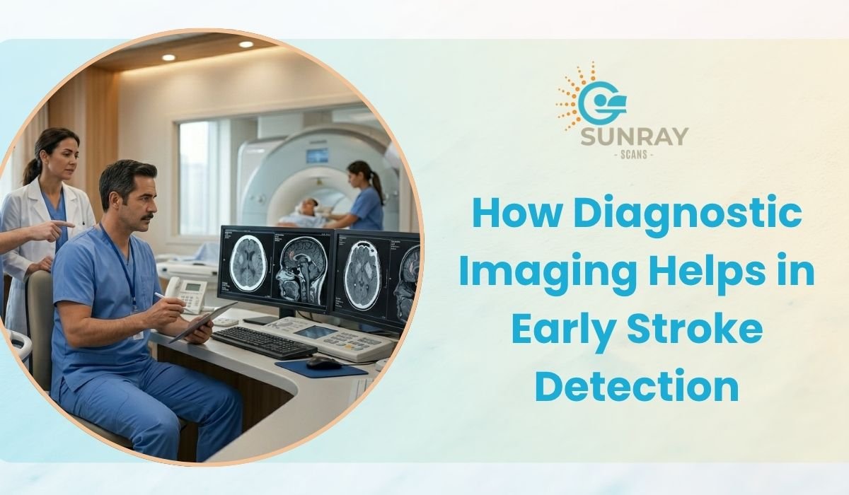
At Sunray Scans, Chennai, access to advanced brain imaging technology, including high-resolution Open MRI and multislice CT, supports timely evaluation when every minute matters. In this blog, we’ll break down how imaging helps detect stroke early, the differences between CT and MRI in stroke care, and why fast, accurate emergency radiology can make the difference between a full recovery and lasting disability.
Why Early Stroke Detection Is So Critical
A stroke occurs when blood flow to part of the brain is interrupted or reduced, depriving brain tissue of oxygen and nutrients. Brain cells begin dying within minutes of this disruption. The longer treatment is delayed, the greater the risk of permanent damage, disability, or death.
There are two main types of stroke:
- Ischemic stroke (about 85% of cases) — caused by a blocked or narrowed artery cutting off blood supply to the brain
- Hemorrhagic stroke — caused by a ruptured blood vessel bleeding into or around the brain
Here’s the critical part: these two types look completely different on the inside, but their symptoms — sudden weakness, slurred speech, facial drooping, confusion, vision problems — can appear nearly identical on the outside. The treatment for one can be actively dangerous for the other. This is exactly why a fast, accurate brain scan isn’t just helpful in stroke care — it’s absolutely essential before any treatment decision is made.
Recognizing Stroke Symptoms Early
While imaging confirms the diagnosis, recognizing symptoms early is what gets a patient to the scanner in time. The widely used F.A.S.T. method helps identify stroke quickly:
- F – Face drooping: One side of the face droops or feels numb
- A – Arm weakness: One arm drifts downward or feels weak when raised
- S – Speech difficulty: Slurred speech or difficulty forming words
- T – Time to call emergency services: Every minute counts
If any of these signs appear, even briefly, getting to a facility equipped for emergency neurological screening immediately is critical — even if symptoms seem to resolve on their own, as this can indicate a transient ischemic attack (TIA), often called a “mini-stroke” and a serious warning sign of a larger stroke risk ahead.
CT Scan: The First Step in Emergency Stroke Imaging
When a patient arrives with suspected stroke symptoms, a CT scan is typically the first imaging test performed, and for good reason — speed.
Why CT Comes First
CT (Computed Tomography) scans are fast, widely available, and excellent at one specific, urgent task: ruling out brain hemorrhage. Since clot-dissolving medication (used for ischemic stroke) can be fatal if given to a patient who’s actually bleeding in the brain, doctors need to rule out hemorrhage almost immediately.
A non-contrast CT scan can typically:
- Detect bleeding in the brain (hemorrhagic stroke) quickly and reliably
- Identify early signs of large ischemic strokes
- Rule out other conditions that mimic stroke, such as brain tumors or abscesses
- Be completed in just a few minutes, which matters enormously in emergency settings
Limitations of CT in Early Stroke Detection
CT does have a notable limitation: in the very early hours of an ischemic stroke, the affected brain tissue may not yet show visible changes on a standard CT scan. This is where more advanced imaging, like CT angiography or MRI, often becomes necessary for a complete picture.
Sunray Scans’ Multislice Advanced CT technology supports faster, high-definition cross-sectional imaging, which assists radiologists in identifying these early or subtle changes more effectively than older CT systems.
MRI for Stroke: Greater Detail for Complex Cases
While CT remains the fast first step, MRI for stroke evaluation offers a level of detail that becomes invaluable, particularly for ischemic strokes in their earlier stages.
How MRI Detects Stroke
MRI uses magnetic fields and radio waves rather than radiation, producing highly detailed images of brain tissue. A specific MRI sequence called diffusion-weighted imaging (DWI) is particularly powerful in stroke diagnosis because it can detect ischemic changes within minutes of stroke onset — often well before changes would be visible on a CT scan.
Key advantages of MRI in stroke evaluation include:
- Detecting small or early ischemic strokes that CT might miss
- Identifying the exact location and extent of affected brain tissue
- Helping distinguish between new and old strokes, which matters for treatment planning
- Assisting in identifying the underlying cause, such as small vessel disease
- Providing more detailed evaluation of the brainstem and cerebellum, areas where CT visualization is often limited
Open MRI: Removing Barriers to Faster Diagnosis
One real-world challenge with MRI, especially in stroke or neurological cases, is patient cooperation. Many patients — particularly those who are anxious, claustrophobic, elderly, or already disoriented from stroke symptoms — struggle inside the narrow, enclosed tunnel of a conventional MRI machine. Restlessness or movement during the scan can compromise image quality at the exact moment precision matters most.
Sunray Scans addresses this with Chennai’s first High-Resolution Open MRI, built using HTS coil technology that delivers sharp, diagnostic-quality brain imaging in a more open, spacious design with quieter operation. For patients undergoing neurological screening who may already be distressed, this more comfortable setup supports better cooperation and, in turn, clearer, more reliable scans.
CT vs MRI for Stroke: Understanding the Difference
Factor | CT Scan | MRI |
Speed | Very fast (minutes) | Takes longer |
Best for | Ruling out hemorrhage, emergency triage | Detecting early/small ischemic stroke |
Radiation | Yes, low dose | None |
Sensitivity for early ischemic stroke | Lower in early hours | Significantly higher |
Brainstem/cerebellum visibility | Limited | Excellent |
Typical use | First scan in emergency setting | Follow-up or detailed evaluation |
In real-world emergency stroke care, these two modalities often work together rather than as alternatives — CT first to rule out bleeding and guide immediate treatment decisions, followed by MRI when more detailed evaluation is needed.
The Role of Vascular Imaging in Stroke Diagnosis
Beyond CT and MRI of the brain itself, evaluating the blood vessels supplying the brain is often a crucial part of a complete stroke diagnosis scan. This is where CT Angiography comes in — a specialized scan that maps blood vessels in detail to identify:
- Blockages or narrowing in arteries supplying the brain
- Aneurysms that may pose a future rupture risk
- Abnormal blood vessel formations
- The overall blood flow pattern to and within the brain
Sunray Scans offers CT Angiography / Peripheral Angiogram imaging, which supports a more complete vascular picture alongside brain imaging — helping doctors understand not just what happened, but why, and what the ongoing risk might be.
Beyond Imaging: Complementary Neurological Tests
While brain imaging forms the core of stroke diagnosis, a complete neurological workup sometimes includes additional tests to assess broader nerve and brain function, especially in cases involving lingering weakness, numbness, or suspected seizure activity following a stroke. Sunray Scans also offers:
- EEG (Electroencephalogram): Maps brain wave activity, useful when seizures are suspected following a stroke
- EMG (Electromyography) & NCS (Nerve Conduction Study): Assesses nerve and muscle function, which can help evaluate weakness that persists after a stroke event
These tests don’t replace brain imaging but often complement it, giving physicians a fuller picture of a patient’s neurological status during recovery and rehabilitation planning.
What to Expect During an Emergency Brain Scan
For patients or family members facing a sudden stroke scare, understanding the process can ease some anxiety:
CT Scan:
- Typically takes just a few minutes
- Patient lies still on a table that moves through the scanner
- Often the very first scan performed in an emergency setting
- May involve a contrast injection for CT angiography
MRI (including Open MRI):
- Takes longer than CT, generally 20–45 minutes depending on the protocol
- No radiation exposure
- Sunray Scans’ open design reduces the closed-in feeling, helping patients remain still for clearer images
- Trained technicians guide patients through the process for added comfort
In genuine stroke emergencies, every step — from symptom recognition to imaging to treatment — is designed to move as quickly as possible. Having a diagnostic center with both rapid CT access and advanced MRI capability under one roof helps streamline this critical window.
Why Fast, Accurate Emergency Radiology Matters
The value of emergency radiology isn’t just in producing an image — it’s in producing the right image, fast, and having it interpreted accurately by an experienced radiologist who can identify subtle but clinically vital findings.
A few factors that genuinely make a difference in time-sensitive neurological imaging:
- Equipment capability: Multislice CT and high-resolution MRI provide clearer images for faster, more confident interpretation
- Radiologist expertise: Subtle early ischemic changes can be easy to miss without trained eyes reviewing the scan
- Reporting turnaround: In stroke care, delayed reports can directly affect treatment timing and outcomes
- Patient comfort and cooperation: Especially relevant in MRI, where motion can compromise image clarity
Sunray Scans supports this with a combination of advanced imaging technology, an experienced radiology team, and one of India’s larger tele-radiology reporting networks — helping ensure scans are reviewed promptly by qualified specialists, including for patients in locations where on-site radiology expertise may be limited.
Stroke Risk Factors Worth Knowing
While this blog focuses on detection through imaging, it’s worth noting the common risk factors that make stroke more likely, since awareness often prompts earlier screening and intervention:
- High blood pressure (the single largest modifiable risk factor)
- Diabetes and poor blood sugar control
- High cholesterol
- Smoking and excessive alcohol consumption
- Atrial fibrillation and other heart rhythm disorders
- Sedentary lifestyle and obesity
- Family history of stroke
For individuals with one or more of these risk factors, periodic neurological screening, even in the absence of symptoms, can sometimes catch warning signs like silent small strokes or vascular narrowing before a major event occurs.
Frequently Asked Questions
Which scan is done first in a suspected stroke — CT or MRI?
CT is almost always performed first in emergency settings because it’s fast and effectively rules out brain hemorrhage, which is critical before deciding on treatment like clot-dissolving medication.
Can a CT scan miss an early stroke?
Yes, in some cases. Very early ischemic strokes may not show visible changes on a standard CT scan within the first few hours, which is why MRI is sometimes used for more detailed evaluation.
Is MRI safe for someone who recently had a stroke?
MRI is generally safe and doesn’t involve radiation. Your treating doctor will determine the right timing and protocol based on the patient’s specific condition and stability.
How long does a brain MRI take?
Depending on the specific sequences required, a brain MRI typically takes between 20 to 45 minutes. Sunray Scans’ Open MRI is designed to make this duration more comfortable for patients.
Can imaging alone diagnose a stroke?
Imaging is central to stroke diagnosis, but it’s interpreted alongside the patient’s symptoms, neurological examination, and sometimes additional tests like EEG or blood work to form a complete clinical picture.
Should someone with stroke risk factors get a screening brain scan even without symptoms?
This is a decision best made with a physician, but for individuals with significant risk factors like uncontrolled hypertension or a strong family history, periodic neurological screening can sometimes be recommended as a precautionary measure.
Final Thoughts
Stroke is one of the few medical emergencies where diagnostic imaging doesn’t just support treatment — it directly determines it. A fast, accurate stroke diagnosis scan can mean the difference between a patient receiving timely, potentially life-saving treatment and losing precious time to uncertainty.
From the rapid clarity of a CT scan to the detailed precision of MRI for stroke evaluation, modern brain imaging has transformed how quickly and confidently doctors can act in critical moments. Combined with complementary neurological screening tools and a strong emergency radiology support system, accurate imaging remains at the heart of effective stroke care.
At Sunray Scans, Royapettah, Chennai, patients have access to advanced Open MRI, multislice CT, CT angiography, and a range of neurological diagnostic tools — all supported by experienced radiologists and a robust tele-radiology network for prompt, reliable reporting when timing matters most.