

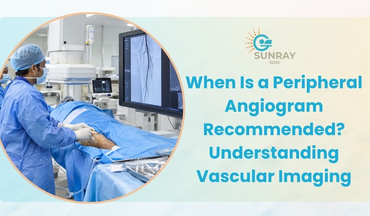
When Is a Peripheral Angiogram Recommended? Understanding Vascular Imaging
When people think about blood vessel problems, their thoughts almost always go straight to the heart. We hear constantly about blocked coronary arteries, heart attacks, and chest pain. Because the heart is so vital, it naturally gets the spotlight.
Your circulatory system is much bigger than just your heart. It is a massive highway network of arteries and veins that stretches for thousands of miles across your entire body. It carries fresh oxygen, sugar, and vital nutrients all the way to your fingertips, down through your digestive organs, and down to the tips of your toes.
When the blood vessels outside of your heart become narrowed, stiff, or blocked, trouble begins in your limbs. You might notice heavy cramping in your calves every time you walk to the market. You might wake up with burning pain in your feet at night. You might notice that a small scrape on your ankle refuses to heal week after week.
Because these symptoms happen in the legs or feet, many people write them off as normal signs of getting older, simple joint stiffness, or mild sciatica.
In reality, these are classic signs of poor circulation. When non-invasive physical checks point toward restricted circulation, doctors turn to a specialized diagnostic test called a peripheral angiogram.
If your healthcare provider has suggested this evaluation, this guide explains what the procedure involves, why it is performed, how modern blood vessel imaging works, and how finding vascular issues early protects your long-term mobility.
What Exactly Is a Peripheral Angiogram?
To understand this test, it helps to break down the medical terminology.
The word “peripheral” refers to the parts of your circulatory system located outside of your heart and brain, mainly focusing on your legs, arms, kidneys, neck, and abdomen. The word “angiogram” comes from two Greek words: “angio,” meaning blood vessel, and “gram,” meaning a drawn record or picture.
A peripheral angiogram is a specialized diagnostic imaging examination that allows doctors to see the inside of your blood vessels with great clarity. It is designed to find out whether blood is flowing smoothly through your limbs or if fatty plaque, calcium deposits, or blood clots are choking off the supply.
During this test, a special liquid called contrast dye is introduced into your bloodstream. Normal blood vessels are soft tissue structures that look invisible or faint on a standard X-ray. The contrast dye absorbs imaging beams, lighting up the entire vascular tree like a bright road map on a screen.
Doctors can spot the exact location, length, and severity of any narrow spot or complete blockage.
The Root Cause: Understanding Peripheral Artery Disease (PAD)
The most common reason doctors recommend peripheral angiography is to diagnose and manage a condition known as Peripheral Artery Disease, or PAD.
PAD develops through a slow process called atherosclerosis. Over many years, cholesterol particles, fatty substances, cellular waste products, and calcium bind together inside the arterial walls. This sticky mixture hardens into a substance called plaque.
As plaque builds up, two problems develop:
- Narrowed Passageways: The inner diameter of the artery shrinks, allowing less blood to pass through.
- Loss of Flexibility: The artery wall becomes stiff and loses its ability to expand when your muscles need extra blood during exercise.
Because your legs are located far away from your heart and contain large muscle groups that demand heavy oxygen during movement, the arteries in the thighs, knees, and calves are usually the first to suffer from restricted blood flow.
Top Warning Signs: When Do Doctors Recommend a Vascular Scan?
Vascular disease rarely starts overnight. It sends quiet warnings that gradually become more noticeable. If you experience any of the following symptoms, your doctor will likely recommend a vascular scan to inspect your circulation:
Intermittent Claudication (Pain While Walking)
This is the most common early sign of vascular trouble in the legs.
- What it feels like: You begin walking, and after a specific distance, a deep, cramping ache or heavy tightness develops in your calf muscles, thighs, or buttocks.
- The telltale pattern: When you stop and sit on a bench to rest for five to ten minutes, the pain completely disappears. But as soon as you resume walking the same distance, the exact same ache returns.
- Why it happens: When resting, your leg muscles need only a trickle of blood to stay comfortable. When walking, those muscles demand four to five times more oxygen. A narrowed artery cannot meet that demand, starving the muscle of oxygen and causing cramp-like pain.
Rest Pain (Burning Pain in the Feet at Night)
If claudication progresses without treatment, the blockage can become so severe that your muscles do not get enough blood even when you are lying completely still.
Patients often experience a burning, throbbing pain across their toes or the balls of their feet, especially when lying flat in bed. Many find themselves dangling their legs over the edge of the mattress or sleeping in an armchair, using gravity to help pull blood down into their feet.
Non-Healing Sores and Ulcers
Your blood carries white blood cells, oxygen, and nutrients needed to repair skin tissue. If you bump your toe or get a small cut that turns into a stubborn sore that will not heal for weeks, it means the skin is not getting enough blood flow to rebuild itself. If ignored, these open sores can turn into infected ulcers or tissue death (gangrene).
Noticeable Temperature and Color Changes in the Legs
Compare your feet and lower legs. Warning signs include:
- One foot feeling icy cold to the touch while the other feels warm.
- Skin on the lower leg looking tight, shiny, and pale, or turning a dark reddish-purple color when dangling down.
- Hair on the calves and toes thinning out or disappearing completely.
- Toenails turning thick, brittle, and growing very slowly.
Sudden Limb Pain (An Acute Emergency)
If an artery suddenly becomes blocked by a floating blood clot, you may experience the “Five Ps”: Pain, Pallor (extreme paleness), Pulselessness, Paresthesia (severe numbness or pins and needles), and Paralysis (inability to move the toes or foot). This is an emergency that requires urgent imaging and hospital care to save the limb.
Who Is at High Risk for Vascular Blockages?
Certain health factors significantly increase the risk of plaque build-up in your peripheral arteries. Doctors have a much lower threshold for ordering a peripheral angiogram in patients with:
- Diabetes: High blood sugar levels accelerate damage to arterial walls, making people with diabetes three to four times more likely to develop peripheral vascular blockages.
- Tobacco Smoking: Smoking damages the smooth inner lining of blood vessels, encourages plaque build-up, and causes arteries to spasm and constrict.
- High Blood Pressure (Hypertension): Constant high pressure batters the arterial walls, creating microscopic tears where cholesterol easily sticks.
- High Cholesterol: High levels of LDL (bad cholesterol) provide the raw material for plaque formation.
- Age Over 50: Vascular stiffness naturally increases as we age.
- History of Heart Disease or Stroke: Atherosclerosis is a widespread condition. If plaque has built up inside your coronary arteries or neck arteries, it is very likely present in your leg arteries as well.
The Diagnostic Path: Steps Before an Angiogram
Doctors rarely jump straight to an angiogram as their very first test. Instead, they follow a stepped evaluation path to assess your circulation safely and comfortably:
Physical Examination and Pulse Checks
Your physician begins by feeling the pulses in your groin, behind your knees, along the inside of your ankles, and on top of your feet. A weak or absent pulse immediately points toward a blockage higher up the leg.
Ankle-Brachial Index (ABI) Test
The ABI is a simple, painless 10-minute test. The technician places blood pressure cuffs on both your arms and both your ankles, using a handheld Doppler wand to listen to blood flow sounds.
By dividing the systolic blood pressure in your ankle by the blood pressure in your arm, the doctor gets a ratio:
- 1.00 to 1.40: Normal, healthy blood flow.
- 0.90 or lower: Points to confirmed Peripheral Artery Disease.
- 0.40 or lower: Points to severe, critical limb ischemia requiring prompt intervention.
Duplex Ultrasound
Before scheduling an angiogram, an ultrasound technician may glide a gel-covered probe over your legs. This uses sound waves to show blood moving through the vessels in real time, confirming which general sections have narrowed.
When the physical exam, ABI, and ultrasound confirm significant arterial narrowing, a detailed angiogram is ordered to create an exact structural map for treatment planning.
The Modern Methods of Blood Vessel Imaging
Today, vascular specialists have three primary ways to perform a peripheral angiogram. The choice depends on your kidney health, overall medical condition, and whether immediate treatment is planned.
CT Angiogram (Computed Tomography Angiography)
A CT angiogram is an advanced, non-invasive method. You do not need surgical incisions or hospital stays.
A small, standard IV line is placed in an arm vein. As contrast dye enters your body, you lie down on a motorized bed that glides through a ring-shaped CT scanner. Within 20 to 30 seconds, the scanner captures thousands of cross-sectional X-ray pictures. Advanced computers assemble these slices into a clean, rotating 3D model of your entire arterial system from your lower back down to your toes.
- Key Advantage: Completely non-invasive, painless, fast, and requires zero recovery downtime.
MR Angiogram (Magnetic Resonance Angiography)
An MR angiogram uses powerful magnetic fields and radio waves instead of X-rays. It is an excellent choice for patients who need to avoid ionizing radiation.
In some cases, modern MR technology can even evaluate blood vessels without needing contrast dye at all, making it a viable alternative for patients with kidney concerns or allergies to traditional iodine dyes.
- Key Advantage: Radiation-free imaging that provides detailed soft tissue and vascular cross-sections.
Catheter-Based Conventional Angiography
This is the traditional, invasive method. It is performed in a specialized hospital operating room called a cardiac catheterization lab.
A vascular surgeon or interventional radiologist numbs your groin with local anesthesia, makes a tiny puncture, and inserts a long, thin, flexible plastic tube called a catheter into the femoral artery. Under live X-ray guidance, the doctor navigates the catheter to the exact spot where the blockage sits and injects contrast dye directly into the vessel.
- Key Advantage: It allows for immediate treatment in the same session. If the doctor finds a tight 90 percent blockage, they can immediately inflate a tiny balloon (angioplasty) and place a metal mesh tube (stent) to open the artery without scheduling a second procedure.
Comparing Diagnostic CT Angiography vs. Catheter Angiography
Feature | Non-Invasive CT Angiogram | Catheter-Based Angiogram |
Invasiveness | Non-invasive (simple arm IV) | Invasive (tube inserted into groin artery) |
Procedure Setting | Outpatient diagnostic scan center | Hospital catheterization lab / operating theater |
Actual Scan Time | Less than 1 minute | 30 to 60 minutes |
Anesthesia Used | None needed | Local anesthesia with light sedation |
Recovery Period | Immediate; walk out and return to work | 4 to 6 hours of strict flat bed rest |
Treatment Capability | Diagnostic mapping only | Can perform ballooning and stenting immediately |
What to Expect: Preparing for Your Peripheral Angiogram
Preparing for a vascular scan is straightforward, but following these basic guidelines ensures a smooth appointment:
- Check Kidney Function: Contrast dye is filtered out of your body by your kidneys through your urine. Your doctor will order a simple blood test (checking serum creatinine and eGFR levels) a few days before the scan to confirm your kidneys are healthy and functioning normally.
- Stay Well Hydrated: Drink plenty of plain water the day before and the day of your scan. Being well hydrated helps protect your kidneys and makes it easier for your body to flush out the contrast dye afterward.
- Fasting Requirements: You will generally be asked not to eat heavy solid foods for four hours prior to the scan, though sipping clear water is usually encouraged.
- Review Your Medications: Bring a complete list of all medications you take. If you take metformin for diabetes, your doctor may ask you to pause it for 48 hours after the scan to protect kidney function. Never stop medications without explicit medical instructions.
- Wear Comfortable Clothing: Choose loose, comfortable cotton clothing without metal buttons, snaps, or zippers around the waist or legs.
What Does the Contrast Dye Feel Like?
When the contrast dye is injected through your IV line during a CT angiogram, it is completely normal to feel a sudden, warm flushing sensation spreading through your chest, belly, and down into your legs. It can sometimes feel like you accidentally passed urine, though you have not. This warm feeling is harmless and disappears completely within 30 to 60 seconds.
How Angiogram Results Shape Your Treatment Plan
A peripheral angiogram is the definitive road map your medical team uses to choose the right treatment. Depending on the severity, length, and location of any blockages discovered, your treatment will fall into one of three categories:
Medical Management and Supervised Walking
If the scan reveals mild to moderate blockages (under 50 to 60 percent narrowing), surgery is rarely needed. Instead, your doctor will focus on aggressive preventive management:
- Prescribing blood thinners (like aspirin or clopidogrel) to prevent blood clots.
- Starting statin medications to stabilize existing plaque and lower cholesterol.
- Enrolling in a structured walking program, which encourages your legs to grow natural detour vessels (collateral circulation) around the narrowed artery.
Endovascular Interventions (Minimally Invasive)
For severe, localized blockages causing difficult walking pain or rest pain, interventional specialists use minimally invasive techniques:
- Balloon Angioplasty: A miniature balloon is inflated inside the narrowed segment to push plaque flat against the vessel walls.
- Stenting: A permanent, expandable metal scaffold is deployed inside the artery to hold it open.
- Atherectomy: A specialized microscopic cutting or laser tool is used to shave down and remove tough, calcified plaque deposits.
Peripheral Bypass Surgery
If an artery is completely blocked over a long distance, or if the vessel is too heavily calcified for a stent, a vascular surgeon may perform a bypass. They take a healthy vein from another part of your leg or use a synthetic graft tube to build a physical bridge around the blocked section, restoring strong, healthy blood flow all the way to your toes.
Why Choose Sunray Scans for Vascular Imaging
When your physician advises an angiogram, you want a diagnostic facility that combines advanced imaging equipment, experienced radiologists, and a comfortable, supportive environment.
For reliable diagnostic care, consider visiting Sunray Scans, a leading medical imaging provider serving patients from their modern center in Royapettah, Chennai.
Sunray Scans provides advanced multi-slice computed tomography (CT) angiography, high-resolution Open MRI systems, and comprehensive ultrasound evaluations under one roof. Their skilled radiographers and senior radiologists ensure every vascular scan is captured with high precision and low radiation exposure, delivering clear, detailed 3D reports that your vascular surgeon or physician can rely on with complete confidence.
What Sets Sunray Scans Apart?
- High-Resolution Multi-Slice CT Technology: Fast, non-invasive imaging that maps complex vascular networks from the abdomen down to the toes in seconds.
- Low-Dose Radiation Protocols: Optimized scanning parameters that protect patient safety while maintaining sharp diagnostic detail.
- Comfort-Focused Care: A welcoming clinical environment designed to keep you relaxed and informed throughout your appointment.
- Comprehensive Diagnostics: Complete diagnostic support, including digital blood testing, ECG, and ultrasound, all available in one facility.
- Prompt, Structured Reports: Quick turnaround times for high-definition 3D reconstructions and radiologist evaluations, keeping your treatment on schedule.
Your legs carry you through every milestone of life. Persistent calf cramps, unexplained foot pain, or cold toes are not things you have to accept as part of aging. They are clear signals that your blood vessels need attention.
Talk with your doctor, take symptoms of poor circulation seriously, and get the answers you need through modern vascular imaging. Finding and treating circulation problems early is the most reliable way to protect your mobility, stay active, and walk through life with confidence.