

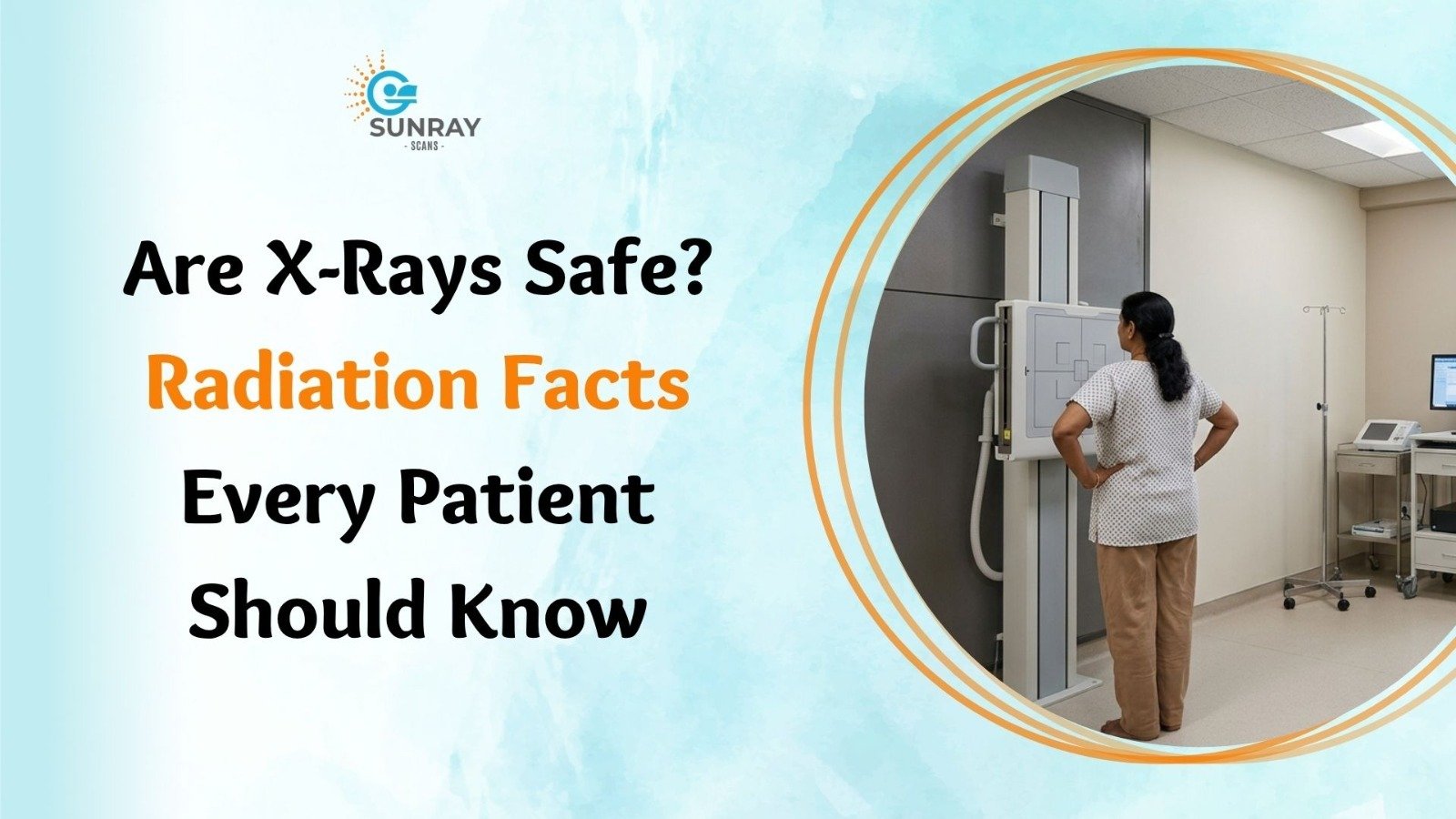
Are X-Rays Safe? Radiation Facts Every Patient Should Know
You trip over a curb, land hard on your wrist, and feel a sharp pain shoot up your arm. Within an hour, you find yourself sitting in an urgent care clinic. The doctor gently presses the swollen bone, nods, and says those familiar words: “Let us get an X-ray to see what is going on.”
For most of us, this is a routine part of modern healthcare. We have seen X-ray machines in hospitals, dental clinics, and airport security lines since childhood.
Yet, the moment the technician places you in front of the cold plate, steps behind a thick protective glass wall, and leaves the room, a quiet flicker of doubt often creeps in. You might look at that heavy barrier and wonder: If this test is completely fine for me, why is the technician hiding behind lead-lined glass? What is that invisible beam doing to my cells? Are X-rays safe, or am I putting my long-term health at risk for a simple bone check?
It is completely natural to have questions about medical radiation. The word “radiation” carries heavy cultural baggage. It makes people think of nuclear power plants, hazardous warning signs, and science-fiction movies.
When you separate sensational headlines from physical facts, the real story of diagnostic imaging is far more reassuring. Medical imaging has evolved dramatically over the past few decades. Today, digital technology allows doctors to capture crystal-clear diagnostic views with only a tiny whisper of the energy once required.
If you or a loved one are preparing for a medical checkup, this guide breaks down the real science behind X-ray radiation exposure, explores what radiation levels mean in daily life, and helps you make informed choices with confidence.
What Actually Is an X-Ray?
To understand whether X-rays are safe, it helps to understand what an X-ray is in the physical world.
An X-ray is simply a form of electromagnetic energy, sitting on the very same spectrum as visible light, radio waves, microwaves, and warmth from the sun. The only real difference between the light coming from your bedside lamp and an X-ray beam is its wavelength and energy level.
Because X-rays have a shorter wavelength and higher energy than visible light, they can pass right through low-density materials like your skin, muscles, and fat.
When those rays hit denser materials, like calcium-rich bones or metal fillings, they are absorbed. A digital detector panel behind your body captures the rays that passed through and the shadows left behind by your bones, creating a detailed structural picture in less than a second.
Why Do People Worry About Radiation?
X-rays are classified as ionizing radiation. This means the energy wave carries enough punch to occasionally knock an electron away from an atom or molecule as it passes through living tissue.
When an electron is displaced, it can temporarily damage cellular structures or DNA molecules inside the cell.
This sounds scary on paper, but your body is not a fragile piece of glass. Living human tissue has evolved over millions of years to repair minor DNA breaks constantly. Every single day, your cells repair thousands of small DNA disruptions caused by normal body heat, metabolic oxygen, and sunlight without any lasting harm.
A danger only arises when the total amount of radiation absorbed over time is exceptionally high, overwhelming the body’s natural cellular repair crews. In diagnostic medicine, the radiation levels used are tiny compared to what it takes to cause harm.
The Great Baseline: Natural Background Radiation
Here is a fact that surprises almost every patient: you are absorbing radiation right now as you read this sentence.
You do not need to visit a hospital or stand near a medical scanner to encounter radiation. We live on a naturally radioactive planet, spinning beneath a massive nuclear furnace called the sun.
Every single day of your life, you are exposed to what scientists call natural background radiation. It comes from several unavoidable natural sources:
- Cosmic Radiation: High-energy particles stream down through our atmosphere from deep space and the sun. If you live at higher elevations, like in the mountains, you absorb more cosmic rays because the air blanket above you is thinner.
- Terrestrial Radiation: The soil, rocks, granite kitchen countertops, and concrete foundations beneath our feet contain trace amounts of natural radioactive elements like uranium, thorium, and radium.
- Radon Gas: A harmless, odorless gas released by the natural decay of minerals in rock and soil, which mixes into the air we breathe every day.
- Internal Radiation in Food: The food you eat contains natural minerals. For example, bananas, potatoes, nuts, and leafy greens are rich in potassium, and a tiny fraction of all potassium on Earth is naturally radioactive Potassium-40. Your own blood and bones emit a faint, harmless background hum of radiation.
Scientists measure the biological impact of radiation in units called millisieverts (mSv).
On average, a typical human being absorbs roughly 2.4 to 3.0 mSv of natural background radiation every single year simply by living on Earth. That works out to about 0.008 mSv every day, completely naturally, without stepping foot in a clinic.
Putting Medical X-Rays in Perspective
When you compare the radiation dose of a routine medical scan to your everyday background exposure, the numbers become far less intimidating.
A standard digital chest X-ray delivers about 0.1 mSv of radiation to your body. That is roughly equivalent to the natural background radiation you absorb from your regular environment over just ten days.
A simple dental X-ray of a single tooth delivers even less: about 0.005 mSv, which is equal to about half a day of normal outdoor living.
Medical Scan / Everyday Event | Typical Radiation Dose (mSv) | Equivalent Natural Background Time |
Dental Bitewing X-Ray (Single) | ~0.005 mSv | About 1 day |
Chest X-Ray (Digital) | ~0.1 mSv | About 10 days |
Airplane Flight (7 to 8 hours) | ~0.03 to 0.05 mSv | About 3 to 5 days of cosmic rays |
Mammogram (Both breasts) | ~0.4 mSv | About 7 weeks |
Annual Background Radiation | ~2.4 to 3.0 mSv | 1 full year of living on Earth |
Abdominal / Pelvis CT Scan | ~7.0 to 10.0 mSv | About 2 to 3 years |
When you look at this comparison, flying in an airplane on a holiday trip exposes you to more cosmic radiation than a routine check of your teeth.
Why? Because cruising at 35,000 feet leaves behind most of the Earth’s protective atmosphere, exposing passengers to an extra sprinkle of space radiation. Yet, few people hesitate before boarding a flight for a family vacation.
Why Does the Technician Step Behind a Barrier?
If a chest X-ray is equal to only ten days of walking in the park, why does the radiographer wear a heavy vest or walk behind a lead-lined wall every single time they press the exposure button?
This is the single most common reason patients feel uneasy, but the answer comes down to occupational frequency.
You might get one or two X-rays in a year, or perhaps a handful over an entire decade. Your total personal exposure from that visit is negligible.
The radiographer, on the other hand, performs thirty, forty, or fifty scans every single day, five days a week, year after year.
If the technician stayed right beside every patient without shielding, those tiny fractions of scattered energy would add up to hundreds of millisieverts over a career. Standing behind lead glass is not a sign that the room is hazardous to you for a two-second visit; it is standard occupational safety to ensure hospital workers do not accumulate high lifetime doses.
Special Considerations: X-Rays During Pregnancy
One of the most sensitive questions in medical imaging is: Can an expectant mother safely get an X-ray?
Whenever a woman is pregnant, doctors treat diagnostic testing with extreme care. Developing babies undergo rapid cell division, making embryonic cells more sensitive to outside influences than adult tissue.
Here are the facts regarding X-ray during pregnancy:
1. Scans Away from the Abdomen Carry Virtually Zero Risk
If an expecting mother twists an ankle, chips a tooth, or develops a stubborn cough, getting an X-ray of the foot, mouth, or chest poses essentially no risk to the baby.
The primary X-ray beam is aimed strictly at the target area (such as the chest or jaw). The amount of scattered radiation that bounces toward the lower abdomen from a chest or dental X-ray is microscopic, well below any threshold known to cause developmental harm.
2. The Golden Rule: Lead Apron Use
When a pregnant patient needs an essential scan, the diagnostic team takes extra precautions by using lead apron use protocols. A heavy, flexible lead-lined blanket or apron is placed over the mother’s abdomen and pelvis. Lead is extremely dense and blocks scattered rays, ensuring the developing baby remains fully shielded.
3. Abdominal and Pelvic Scans
If an emergency requires an X-ray directly over the belly or pelvis, doctors always weigh the benefit against the risk. Non-radiation alternatives, such as diagnostic ultrasound or an MRI, are always considered first.
If an X-ray remains clinically necessary to treat a serious maternal condition, your physician will consult with a medical physicist to keep the exposure settings at the lowest possible level.
Always inform your doctor and technician if you are pregnant or think you might be before any scan begins.
Protecting Little Ones: X-Ray Safety for Kids
Parents naturally feel protective when a pediatrician recommends an imaging check for a toddler or young child. Children have growing bodies with rapidly dividing cells, and because they have a full lifetime ahead of them, medical guidelines treat pediatric radiation with great care.
To ensure complete X-ray safety for kids, modern diagnostic centers follow an internationally recognized medical standard known as the ALARA Principle.
What Is the ALARA Principle?
ALARA stands for As Low As Reasonably Achievable. It means medical teams must use the absolute minimum amount of radiation necessary to capture a sharp, medically useful image, and not a fraction more.
In practical pediatric care, this means:
- Child-Sized Dosing: Technicians manually adjust the machine’s power settings based on the child’s exact weight and height. A five-year-old child receives only a fraction of the beam energy used for a grown adult.
- Narrow Beam Collimation: The machine uses lead shutters to narrow the beam so it shines only on the exact bone in question, preventing surrounding areas from receiving unnecessary exposure.
- Rapid Digital Exposures: Modern digital sensors capture pictures in milliseconds, meaning kids do not have to hold still for long, reducing the need for repeat shots due to wriggling.
When your child has a suspected bone break, a swallowed coin, or a severe chest rattle, the real medical danger comes from missing a serious condition, not from the tiny, controlled dose of a pediatric X-ray.
How Often Can You Get X-Rays?
Patients who manage chronic health issues often ask: Is there a strict limit on how often can you get X-rays?
There is no arbitrary number or annual quota that says you can only have three, five, or ten X-rays in a single year. Medical decisions are never based on a scorecard; they are based on a risk-versus-benefit balance.
Every time a doctor considers ordering a scan, they ask two simple questions:
- Will the information from this scan directly help treat or heal the patient?
- Does the medical risk of NOT doing the scan outweigh the tiny physical risk of the radiation?
If you have a broken leg that is being set with metal pins, your orthopedic surgeon might need an X-ray before the procedure, one during alignment, another right after surgery, and follow-up checks at two, six, and twelve weeks.
While that might sound like a lot of pictures, each scan provides crucial information to confirm that the bone is knitting straight and not developing an infection.
The health danger of a crooked, improperly healed bone or an undiagnosed internal infection is immediate, serious, and life-altering. The theoretical risk from those small, spaced-out digital X-rays is practically zero.
4 Practical Steps to Minimize Your Radiation Exposure
While modern imaging is exceptionally safe, taking a smart, proactive approach to your medical records is always good practice:
- Keep a Personal Imaging History: Maintain a digital folder or notes app list of your past scans, including dates, clinics, and body parts examined. If you change doctors or visit a new specialist, bring your previous digital reports so they do not repeat a test that was already done last week.
- Ask Thoughtful Questions: It is always fine to ask your physician: “How will this test change my treatment plan?” or “Could an ultrasound or MRI give you the same answers without radiation?” A good doctor will gladly explain why a specific tool is best.
- Hold Still During the Scan: The most common reason a scan has to be repeated is motion blur. When the technician asks you to hold your breath and stay completely still for two seconds, following instructions carefully ensures they get a perfect shot on the very first try.
- Choose Modern Digital Facilities: Older film-based X-ray systems required significantly higher radiation doses to expose chemical sheets. Always choose centers that use modern high-frequency digital radiography equipment.
Why Quality and Safety Matter in Diagnostic Imaging
When you need an X-ray for yourself or your family, you want to know that the facility handling your care uses modern, calibrated equipment operated by certified professionals who put safety first.
For dependable diagnostic care in Tamil Nadu, consider visiting Sunray Scans, a leading medical imaging provider serving patients from their modern center in Royapettah, Chennai.
Sunray Scans utilizes modern low-dose digital radiography systems alongside a complete range of diagnostic services, including high-resolution Open MRI, multi-slice CT angiography, advanced ultrasound, and automated lab testing. Their experienced team of radiographers and senior radiologists follows strict international radiation protection guidelines, ensuring every scan delivers sharp diagnostic clarity with minimal exposure.
Why Patients Trust Sunray Scans
- Modern Low-Dose Digital Equipment: Advanced digital sensor panels that cut radiation requirements by up to 70 to 80 percent compared to older traditional systems.
- Strict Safety and Shielding Protocols: Dedicated lead aprons, thyroid shields, and pediatric dose adjustments for complete peace of mind.
- Gentle, Patient-Focused Care: Experienced technicians who guide children, seniors, and anxious patients with patience and care.
- Fast Digital Reports: Instant image previews and quick report delivery so you can consult your treating physician without delay.
Medical imaging is one of the most powerful diagnostic tools ever created. It takes the guesswork out of healthcare, allows doctors to catch infections early, and helps broken bones heal straight and strong.
When performed on modern digital equipment with sensible precautions, diagnostic X-rays are safe, gentle, and an indispensable partner in protecting your long-term health.
Talk with your doctor, ask questions whenever you feel unsure, and get the answers you need to keep living your life in good health.