

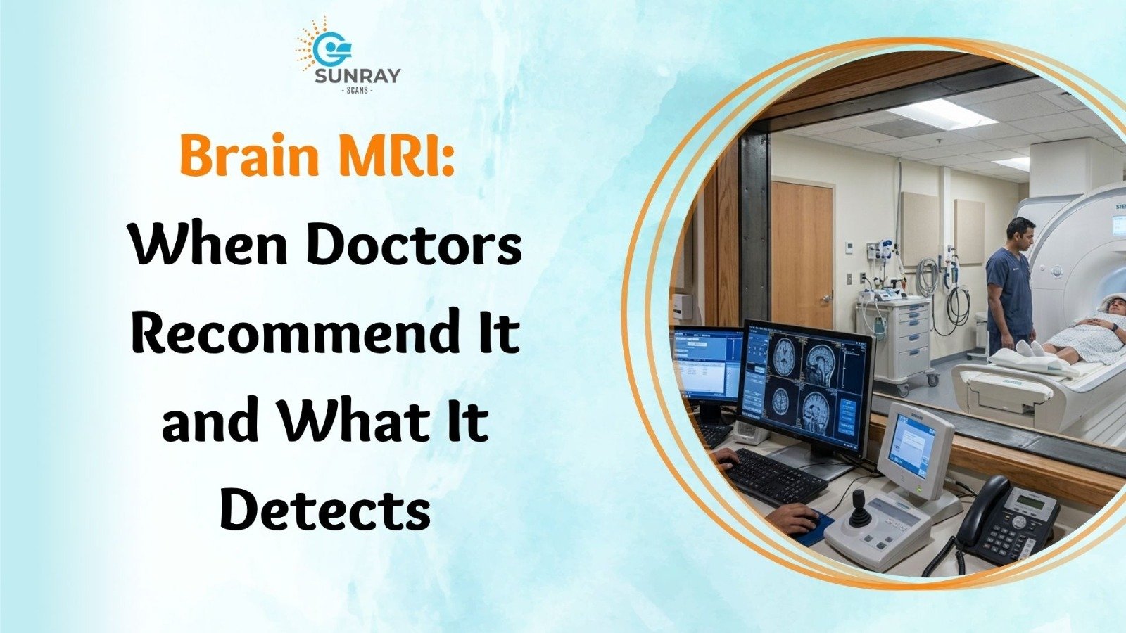
Brain MRI: When Doctors Recommend It and What It Detects
When you visit a clinic with a stubborn health issue, you usually expect a straightforward checkup. A sore throat gets a quick swab. A sprained ankle gets an ice pack and an elastic wrap.
When symptoms start happening inside your head, things feel very different.
A new kind of throbbing headache keeps you awake at night. A sudden wave of dizziness causes you to reach for a wall to stay upright. A family member notices that your speech sounds slightly slurred, or you experience a sudden, frightening blackout that you cannot remember.
In these moments, it is completely normal for fear to set in. Your mind immediately races through worst-case possibilities. You want quick, clear answers, and your doctor knows that guessing is never an option when it comes to the nervous system.
This is why neurologists and physicians turn to a brain MRI.
Magnetic Resonance Imaging is one of the greatest achievements in modern healthcare. It allows doctors to look through the solid bone of the skull and inspect the living brain in breathtaking detail, all without making a single cut or using harmful radiation.
If your doctor has recommended this scan, or if you are trying to understand why this test is so widely trusted, this comprehensive guide explains how it works, when doctors order it, what it detects, and what you can expect during your appointment.
What Exactly Is a Brain MRI?
To understand why a magnetic scan is so effective, it helps to understand what makes your brain unique.
Your brain is a soft, delicate organ made largely of water, healthy fats, blood vessels, and billions of interconnected nerve cells. Traditional imaging tools like basic X-rays work well for solid bones, but they struggle with soft tissues. On an X-ray, the thick bone of your skull absorbs the energy, leaving the soft brain hidden in a vague shadow.
A brain MRI solves this problem by using physics rather than radiation.
The machine uses three safe, natural elements:
- A powerful magnetic field.
- Gentle radio waves.
- High-speed computer processors.
The Science Behind the Image
Inside your body, every single water molecule contains hydrogen atoms. Under normal conditions, these tiny atoms spin in random, unorganized directions.
When you lie down inside an MRI machine, the scanner’s powerful magnetic field causes these hydrogen atoms to line up in the same direction, like compass needles pointing north.
Next, the machine sends a quick burst of harmless radio waves through your head. This radio wave gently knocks the atoms out of alignment.
When the radio signal is turned off, the atoms relax back to their original position. As they snap back into place, they release a faint burst of radio signals of their own.
Because different types of brain tissue (such as grey matter, white matter, fluid, and blood) contain different amounts of water, their atoms relax at different speeds. Sensitive antenna coils wrapped around your head pick up these tiny signals. A computer reads millions of these signals every second and paints a three-dimensional map of your brain, showing fine cross-sections as thin as a single millimeter.
Brain MRI vs. Brain CT Scan: What Is the Difference?
Patients frequently ask why a doctor chooses an MRI instead of a Computed Tomography (CT) scan, especially since CT scans are faster. Both tools are valuable, but they serve different clinical purposes.
Feature | Brain CT Scan | Brain MRI |
Technology Used | Rotating X-ray beams (radiation) | Magnetic fields and radio waves (zero radiation) |
Scan Duration | Very fast (3 to 5 minutes) | More thorough (15 to 45 minutes) |
Best Used For | Fresh bleeding, skull bone fractures, urgent trauma | Soft tissue detail, small nerve paths, early diseases |
Soft Tissue Contrast | Moderate | Exceptional |
Safety Profile | Uses ionizing radiation | Completely radiation-free |
In an emergency room setting, if someone arrives after a severe car crash or a sudden head injury, doctors usually order a CT scan first. It takes only a couple of minutes and quickly rules out cracked bones or large pools of fresh blood.
When doctors need to evaluate subtle nerve inflammation, identify tiny areas of tissue loss, check for early infections, or examine deep brain structures, an MRI is the undisputed gold standard.
When Do Doctors Recommend a Brain MRI?
Neurologists, general physicians, and surgeons recommend a magnetic brain scan for many clinical reasons. Here are the most common situations where this test provides essential answers:
1. Investigating Persistent or Unusual Headaches
Almost everyone gets a headache from time to time due to work stress, lack of sleep, or dehydration. These common aches usually fade with rest or simple over-the-counter medicine.
However, doctors pay close attention when headache patterns change drastically. Getting an MRI for headache symptoms is recommended when you experience:
- A “thunderclap headache,” which is an excruciating pain that peaks within sixty seconds.
- Headaches that wake you from a sound sleep in the middle of the night.
- Head pain that consistently worsens when you cough, sneeze, or bend forward.
- New, daily headaches that begin for the very first time after age fifty.
- Headaches accompanied by stiff neck muscles, persistent nausea, or a fever.
- Headaches that come with vision loss, tingling, or weakness in your arms or legs.
An MRI helps rule out increased pressure inside the skull, swollen blood vessels, structural blockages of spinal fluid, and underlying growths.
2. Urgent Evaluation: MRI for Stroke Symptoms
A stroke happens when blood flow to a section of your brain is interrupted. Without oxygen, brain cells begin to die within minutes.
When evaluating a patient with sudden weakness, facial drooping, or speech trouble, doctors often rely on an MRI for stroke assessment, specifically using an advanced technique called Diffusion-Weighted Imaging (DWI).
Diffusion MRI tracks the microscopic movement of water molecules between cells. When brain cells are starved of oxygen, their outer walls malfunction and water becomes trapped inside. A diffusion scan can spot this trapped water within three to five minutes of a stroke starting, long before standard CT scans can detect any tissue changes.
This early detection window allows doctors to administer clot-busting medications or perform emergency procedures to clear the blocked artery and save healthy brain tissue.
3. Evaluating Seizures: MRI for Epilepsy
A seizure occurs when groups of brain cells produce sudden, abnormal surges of electrical activity. While an EEG (electroencephalogram) records the live electrical waves, an MRI for epilepsy searches for the physical, structural trigger behind those electrical misfires.
A specialized high-resolution scan can uncover:
- Mesial Temporal Sclerosis: A tiny patch of scar tissue in the hippocampus, which is the most common cause of focal seizures.
- Cortical Dysplasia: Small areas where brain cells did not form smooth layers during early childhood development.
- Cavernous Malformations: Tiny clusters of abnormal, fragile blood vessels that leak microscopic drops of blood and irritate nearby nerves.
Finding the exact physical spot causing seizures helps neurologists select the most effective medications or evaluate whether minimally invasive surgery can cure the condition completely.
4. Detecting Brain Tumours and Cysts
One of the most critical roles of imaging is brain tumour detection.
Brain growths can be benign (non-cancerous, slow-growing masses like meningiomas or acoustic neuromas) or malignant (aggressive cancers like glioblastomas or metastatic tumors that spread from other organs).
An MRI provides unmatched clarity for tumor evaluation:
- It marks the precise boundary between a tumor and healthy brain tissue.
- It shows whether surrounding brain tissue is swollen with fluid (edema).
- It checks whether the mass is pressing on vital nerve pathways or shifting the midline of the brain.
- It guides neurosurgeons during operations, acting like a GPS map so they can remove the growth without disturbing critical speech or motor centers.
Specialized Brain MRI Techniques
MRI technology is not a single, rigid test. Doctors can run specialized imaging sequences depending on the exact question they need to answer:
1. Contrast-Enhanced MRI
For certain scans, a nurse or technician will inject a special liquid called gadolinium contrast into a vein in your arm. Gadolinium is a safe, non-iodine material that temporarily alters the magnetic properties of nearby tissue.
Healthy brain tissue has a protective shield called the blood-brain barrier, which prevents foreign substances from entering. Many tumors, active infections, and inflamed areas have leaky blood vessels that break this barrier. The contrast dye collects inside these problem areas, causing them to light up bright white on the scan screen.
2. Magnetic Resonance Angiography (MRA) and Venography (MRV)
If your doctor wants to inspect your blood vessels without threading long tubes through your body, they order an MRA or MRV.
These scans adjust magnetic settings to make static brain tissue look invisible while highlighting moving blood. An MRA checks the arteries for balloon-like weak spots (aneurysms), narrow sections, or dangerous tangles of vessels called arteriovenous malformations (AVMs). An MRV checks the large veins for dangerous blood clots (cerebral venous thrombosis).
3. Functional MRI (fMRI)
While a standard scan takes detailed pictures of brain anatomy, a functional MRI watches the brain in action.
When you use a specific part of your brain, like wiggling your fingers, reading a sentence, or listening to music, that specific region demands extra oxygen. The fMRI detects tiny shifts in blood oxygen levels as you perform simple tasks inside the scanner.
Surgeons use fMRI maps before complex brain operations. By identifying the exact coordinates of your personal speech and movement centers, the surgical team can plan safe pathways that protect your vital abilities.
4. MR Spectroscopy (MRS)
Think of MRS as a virtual tissue biopsy without needles. Instead of producing an anatomical picture, it produces a graph showing the chemical makeup of a suspicious spot.
By measuring the ratios of natural brain chemicals like choline, creatine, and N-acetylaspartate, an MRS helps doctors distinguish between a recurring tumor, an old infection, or harmless scar tissue.
Summary: What Can a Brain MRI Detect?
Medical Category | Specific Conditions Identified |
Vascular Disorders | Ischemic strokes, brain aneurysms, bleeding, venous clots, AVMs |
Abnormal Growths | Benign meningiomas, acoustic neuromas, pituitary adenomas, malignant cancers |
Demyelinating Diseases | Multiple Sclerosis (MS) plaques, optic neuritis |
Infections | Brain abscesses, encephalitis, meningitis, fungal and parasitic cysts |
Degenerative Conditions | Alzheimer’s disease, vascular dementia, regional brain shrinkage |
Structural & Congenital | Chiari malformations, hydrocephalus (excess fluid), arachnoid cysts |
Sensory Nerve Issues | Trigeminal neuralgia (facial nerve pain), inner ear balance nerve compression |
The Patient Experience: What to Expect During Your Scan
Knowing what happens during an appointment helps remove anxiety and lets you approach your test with total confidence.
Step 1: Preparation and Safety Checks
Before entering the scan room, you will complete a thorough safety questionnaire. Because the MRI machine is a massive, permanent magnet, you must remove all metallic items from your body:
- Watches, jewelry, eyeglasses, and hairpins.
- Hearing aids, dentures, and metallic dental plates.
- Belts, wallets, credit cards (the magnet can erase magnetic strips), and mobile phones.
You will usually change into comfortable, clean clinic cotton scrubs or a gown to ensure no hidden metal threads, snaps, or zippers interfere with the magnetic field.
Step 2: Settling Onto the Table
You will lie down on your back on a cushioned, motorized examination bed.
The technologist will place a soft, helmet-like plastic frame over your head. This frame is called a head coil. It does not touch your face tightly; it acts as a sensitive radio antenna to capture the clearest possible signals from your brain.
The technician will tuck soft foam pads around your head to keep you comfortable and help you avoid accidental movement.
Step 3: Inside the Scanner
The motorized bed will slowly glide into the scanner. The inside of the machine is well-lit, open at both ends, and features a continuous stream of fresh air blowing gently across your face.
If you are having your scan on a modern open-sided machine, the sides remain completely wide open to the room, eliminating any feeling of confinement.
Step 4: The Sounds of the Scan
The scan itself is completely painless. You will not feel the magnetic field or the radio waves.
You will, however, hear distinctive sounds. As the internal magnetic coils turn on and off, the machine produces rhythmic knocking, thumping, buzzing, and clicking noises.
The clinic will provide you with soft foam earplugs or comfortable headphones playing relaxing music to soften the noise.
Step 5: Staying Still
The most important job you have during the test is to stay as still as possible.
Each imaging sequence lasts between two and five minutes, with short quiet pauses in between. Even small movements, like nodding, turning your head, or clenching your jaw, can blur the image, much like a camera taking a picture of a moving car.
If an image is blurred, that sequence must be repeated, making the appointment longer. Closing your eyes, breathing slowly, and focusing on relaxing thoughts makes the time pass quickly.
Pro Tip: The Emergency Squeeze Ball
The technologist sits right outside the room behind a large clear observation window, watching you constantly and communicating through a two-way speaker system. Before the scan starts, they will place a small rubber squeeze ball in your hand. If you feel uncomfortable, need to cough, or want to pause at any moment, simply squeeze the ball. The scan pauses instantly, and the technician will assist you right away.
Overcoming Scan Anxiety and Claustrophobia
For many people, the hardest part of an MRI is not the test itself, but the fear of enclosed spaces. If you experience claustrophobia, you are far from alone. Millions of people feel nervous at the thought of lying inside a traditional tube scanner.
Fortunately, modern diagnostic care has evolved to put patient comfort first:
- Open MRI Options: Advanced diagnostic centers now offer open MRI scanners. Instead of an enclosed cylinder, an open machine features a top plate and a bottom plate with wide open sides. You can see the entire room, look at the technologist, and enjoy open air throughout your scan.
- Companion Support: In open imaging suites, a family member or friend can often sit right beside you and hold your hand during the test.
- Mindful Breathing: Practicing slow, four-second inhales and four-second exhales keeps your nervous system in a restful state.
- Open Communication: Let the clinic staff know in advance if you feel nervous. Experienced teams know how to guide you with care, taking breaks between sequences so you never feel rushed.
How to Prepare for Your Brain MRI
Getting ready for a brain scan requires very little effort:
- Food and Drink: For a standard non-contrast scan, you can eat, drink, and take your regular daily medications normally. If your doctor has ordered a contrast-enhanced scan, you may be asked to avoid heavy meals for two to three hours before your appointment.
- Kidney Health Checks: If contrast dye is required, your doctor will review a recent simple blood test (serum creatinine) to confirm that your kidneys are functioning well and will easily filter the contrast material out through your urine.
- Disclose Metal Implants: Always tell your team if you have surgical clips, metallic plates, artificial heart valves, joint replacements, or modern pacemakers. Most modern implants are made of titanium and are completely safe, but the team will verify your medical device card to set the correct machine parameters.
- Skip Eye Makeup: Some cosmetic products, particularly dark mascaras and metallic eye shadows, contain trace mineral pigments that can cause minor magnetic interference and blur images around the eyes and frontal lobes. Arriving with a clean face is best.
Understanding Your Brain MRI Results
After your scan is complete, the collected data is processed into hundreds of detailed digital images.
A medical doctor who specializes in reading diagnostic images, known as a radiologist, reviews every sequence slice by slice. They inspect the symmetry of both brain hemispheres, examine the fluid-filled spaces, check blood vessel pathways, and evaluate tissue textures.
The radiologist writes a structured medical report detailing their observations:
- Normal Findings: The brain structures show expected size, shape, and position for your age. There are no signs of bleeding, stroke, abnormal masses, or fluid buildup.
- Incidental Findings: Very often, an MRI discovers tiny, harmless variations that have nothing to do with your symptoms, like an innocent fluid cyst or minor age-related white matter spots. Your doctor will explain which findings matter and which are simply normal anatomical quirks.
- Diagnostic Findings: If an issue is present, such as an inflamed nerve, a narrowed vessel, or a tumor, the report describes its exact location, size, and characteristics, giving your treating physician the exact data needed to create a tailored treatment plan.
Why Choose Sunray Scans for Your Brain MRI
When you or a loved one needs a neurological evaluation, you want a diagnostic facility that balances world-class technical precision with compassionate, patient-first care.
For outstanding imaging services, consider visiting Sunray Scans, a leading medical diagnostic provider serving patients from their modern center in Royapettah, Chennai.
Sunray Scans proudly introduced Chennai’s first-ever High-Resolution Open MRI system, designed specifically to eliminate the fear of tight spaces while delivering pristine diagnostic detail. Their modern facility combines advanced soft-tissue imaging protocols with a quiet, stress-free clinical environment. Every scan is conducted by certified, caring radiographers and interpreted by experienced senior radiologists, ensuring you receive clear, dependable answers without delay.
What Sets Sunray Scans Apart?
- True High-Resolution Open MRI: Experience wide-open sides, silent scanning technology, and complete peace of mind without the claustrophobia of traditional closed tubes.
- Advanced Neurological Protocols: Specialized imaging sequences for stroke screening, chronic headaches, epilepsy mapping, and vascular anomalies.
- Comfort-First Patient Care: Courteous staff who take the time to answer your questions, accommodate family companions, and guide you through every step.
- Accurate Reporting: Fast report turnaround times so you can take your digital images and structured reports directly to your neurologist or physician.
- Accessible and Transparent Care: High-quality healthcare delivered at fair, competitive rates for patients across Chennai.
Your brain controls every thought, movement, memory, and emotion that makes you who you are. When persistent symptoms signal that something is wrong, guessing only prolongs anxiety and delays relief. A modern brain scan gives your medical team the clear, safe, and accurate view they need to protect your long-term health.
Talk with your doctor, take your warning signs seriously, and schedule your evaluation with a trusted imaging partner to get the clarity and peace of mind you deserve.