

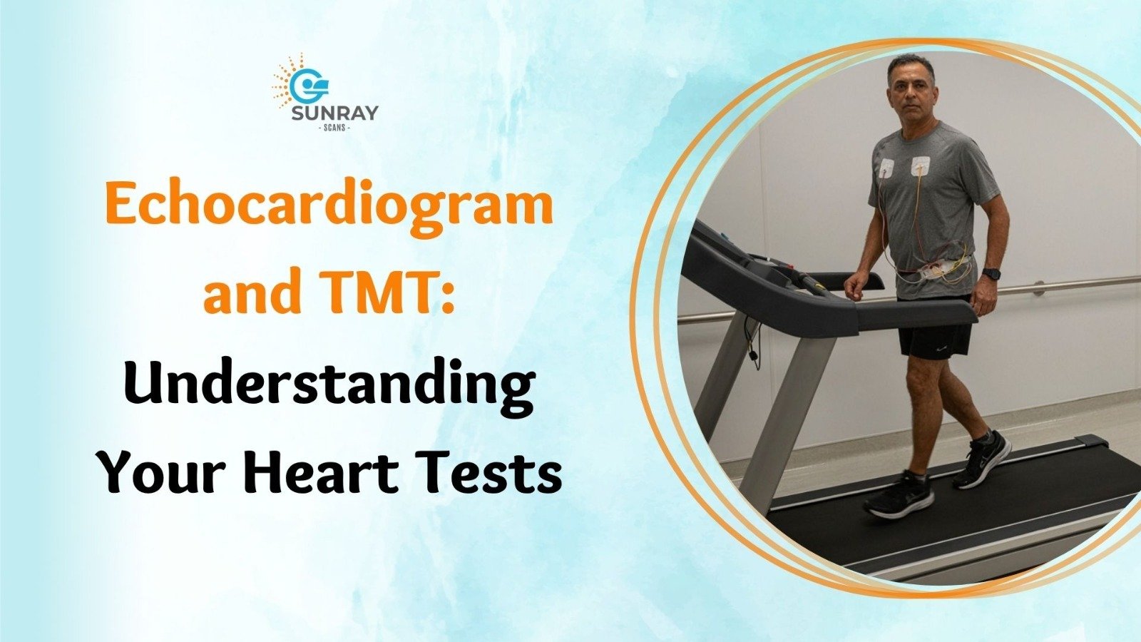
Echocardiogram and TMT: Understanding Your Heart Tests
You wake up, climb a flight of stairs, carry bags of groceries inside, or rush to catch a bus, and you expect your body to keep pace without protest. We rarely stop to think about the steady pump working quietly inside our chest.
Day in and day out, your heart contracts roughly one hundred thousand times, circulating thousands of liters of blood through your body without taking a single break.
When something feels slightly off, it commands your full attention. Perhaps you notice a sudden flutter in your chest while sitting on the sofa. Maybe you feel winded after a short walk down the street, or your doctor hears a soft clicking sound when listening through a stethoscope during a regular physical.
In that moment, worry tends to set in quickly. When a cardiologist hands you a prescription requesting an echocardiogram alongside a TMT test, questions naturally follow.
Why would you need two different evaluations for the same organ? Does one test replace the other? How do they work, and what do they show?
Your heart is a complex machine that relies on electrical signals, muscular pumping chambers, one-way valves, and vascular fuel lines. Because of this multi-layered design, no single test tells the whole story.
This guide breaks down the science behind an echocardiogram and a treadmill test in simple, everyday language. It explains how both tools work together, what you can expect during each appointment, and how these tests help protect your cardiovascular health.
The Heart as a House: Electricity vs. Plumbing vs. Structure
To understand why doctors order different heart function tests, it helps to picture your heart as a well-built house.
A house needs three primary systems to work properly:
- The Electrical Wiring: Copper wires running through the walls that send power to switch on the lights and appliances.
- The Structural Walls and Doors: Solid foundation walls dividing rooms, with swinging doors that open and close to control movement from one room to the next.
- The Plumbing Pipes: Clean water pipes that deliver water throughout the house and drain lines that carry waste away.
- An Electrocardiogram (ECG) measures the electrical wiring. It looks at the rhythm of the electrical impulses that tell the heart muscles when to beat.
- An Echocardiogram evaluates the physical structure and swinging doors. It looks at the heart muscle walls, measures how wide the chambers expand, and watches the heart valves open and shut.
- A Treadmill Test (TMT) tests the plumbing pipes under pressure. It monitors how well the coronary arteries deliver blood when the engine is pushed to work harder.
If your doctor suspects an issue with the doors (valves) or walls (pumping muscle), they need an ultrasound view. If they suspect narrowed supply lines (arteries), they need to watch how the heart performs under physical effort.
What Is an Echocardiogram?
An echocardiogram, often called an “echo” or a cardiac ultrasound, is a safe, painless, non-invasive diagnostic procedure that uses high-frequency sound waves to create moving, real-time pictures of your heart.
It works on the exact same ultrasound technology that obstetricians use to look at a developing baby in the womb.
There are no X-rays, no cutting, and zero radiation involved. Instead, a handheld wand called a transducer probe sends high-frequency sound waves into your chest.
These sound waves travel through skin and ribs, bouncing off the dense heart muscle walls, delicate valve flaps, and moving blood cells. The probe captures those returning sound echoes, and a specialized computer translates them into live, moving video on a screen.
What an Echocardiogram Shows
When a cardiologist looks at an echocardiogram, they are evaluating several key mechanical features:
- Size and Shape of the Heart: It measures the exact thickness of the muscular walls and the volume of all four chambers (atria and ventricles). If high blood pressure has caused the muscle to become abnormally thick and stiff (hypertrophy), the scan shows it clearly.
- Pumping Strength (Ejection Fraction): One of the most important numbers on an echo report is the Ejection Fraction (EF). This is the percentage of blood pumped out of the main pumping chamber (left ventricle) with each heartbeat. A normal EF typically sits between 55 percent and 70 percent. A lower number indicates that the heart muscle is weakened, which can be an early sign of heart failure.
- Heart Valve Function: Your heart contains four vital valves: tricuspid, pulmonary, mitral, and aortic. These act like one-way swinging doors. An echo shows whether a valve is stiff and narrow (stenosis), preventing blood from moving forward, or loose and leaky (regurgitation), allowing blood to wash backward.
- Pericardial Fluid: It checks the outer protective sac surrounding the heart (pericardium) to confirm there is no excess fluid buildup causing pressure on the muscle.
- Blood Clots or Masses: It verifies that no stagnant blood clots have formed inside the chambers, which is especially important for patients who have irregular heart rhythms like atrial fibrillation.
The 2D Echo Procedure: What to Expect During Your Appointment
Knowing the step-by-step 2D echo procedure helps you approach your test with complete peace of mind.
Step 1: Getting Settled
When you arrive at the diagnostic center, you will be shown to a private, softly lit ultrasound room. You will be asked to remove your upper clothing and put on a comfortable clinic gown that opens at the front.
Step 2: Electrode Placement
You will lie down on a padded examination bed, usually turned onto your left side. Lying on your left side allows gravity to bring your heart closer to the front wall of your chest, giving the ultrasound wand the clearest possible view through your ribs.
The technician will place three small, sticky sensor patches on your chest to record your resting ECG simultaneously. This helps the computer match the ultrasound video frames with each phase of your heartbeat.
Step 3: The Scanning Process
The technician applies a clear, water-based gel to your chest. The gel might feel slightly cool for a few seconds. Its purpose is simple: it eliminates any air pockets between your skin and the probe, allowing sound waves to travel smoothly into your body.
The technician will gently glide the probe across different areas of your chest:
- Along the left edge of your breastbone.
- Just below your left nipple.
- Slightly above your stomach at the base of your ribs.
- At the base of your throat near your collarbone.
Step 4: The Sounds of the Scan
During the scan, you will suddenly hear a rhythmic, loud whooshing sound coming from the machine.
Do not be alarmed. This is the Colour Doppler function turning on. The machine converts the sound of blood flowing through your heart valves into an audible signal and a color-coded map on the screen.
Red shows blood flowing in one direction, while blue shows blood moving the other way. This allows the doctor to spot small leaks across your valves with high precision.
You may be asked to hold your breath for a few seconds or shift positions slightly. The entire scan takes between 20 and 40 minutes. Once done, the gel is wiped away with a towel, you put your clothes back on, and you are free to return to your normal day with zero restrictions.
What Is a TMT Test (Treadmill Stress Test)?
While an echocardiogram inspects the physical layout of the heart while you rest comfortably on a bed, a TMT test evaluates how your cardiovascular system behaves when pushed to work harder.
When you are resting, your heart pumps about four to five liters of blood per minute. When you walk briskly or climb a hill, your leg muscles demand significantly more oxygen. Your heart responds by beating faster and pumping up to four or five times more blood per minute.
If your coronary arteries (the blood vessels that supply the heart muscle itself) have mild or moderate plaque buildup, you might feel completely fine while sitting down. Enough blood trickles past the narrow spots to satisfy the heart’s resting needs.
Once you begin walking up an incline on a treadmill, those narrowed arteries cannot keep up with the surging demand for oxygen. The heart muscle becomes starved of fuel, which produces distinct electrical changes on an ECG monitor and causes symptoms like chest heaviness or breathlessness.
A TMT test creates a controlled, safe environment to evaluate this stress response step by step.
The TMT Test Procedure: Step by Step
Understanding how a treadmill test works ensures you arrive prepared:
- Skin Preparation: Small areas on your chest are cleaned so ten sticky electrode patches adhere firmly to your skin. These wires connect to a digital ECG machine. A blood pressure cuff is placed around your arm.
- Resting Baseline: The medical team records your resting blood pressure and resting ECG while you are lying down, sitting, and standing up.
- The Bruce Protocol: You step onto a motorized treadmill. The test begins at a slow, gentle walking speed (around 2.7 km/h) with a mild 10 percent incline. Every three minutes, the speed and incline increase slightly.
- Continuous Monitoring: The technician and supervising doctor watch your live ECG patterns on a screen, track your pulse rate, and take your blood pressure at regular intervals.
- Reaching the Target: The walk continues until your heart rate reaches its calculated target, which is roughly 85 percent of your age-predicted maximum heart rate, or until you feel too tired to continue safely.
- Recovery Observation: After the treadmill stops, you immediately sit down while the team continues monitoring your ECG and blood pressure for another five to eight minutes to watch how quickly your heart returns to its normal baseline.
TMT vs ECG: Clearing Up the Confusion
A common point of confusion for patients is the difference between a routine resting ECG and a TMT test. Both use sticky chest patches, and both print out wavy electrical lines on graph paper.
Why do a TMT vs ECG comparison if they look so similar?
Diagnostic Feature | Standard Resting ECG | Treadmill Stress Test (TMT) |
Patient Position | Lying completely flat on a bed | Walking/jogging on an inclined treadmill |
Duration of Test | About 1 to 2 minutes | 15 to 30 minutes total (6 to 12 minutes walking) |
Heart Workload | Resting state (60 to 90 beats/min) | High physical effort (up to 140 to 170 beats/min) |
Primary Goal | Spots past heart attacks or resting rhythm flaws | Reveals hidden coronary artery blockages |
Limitation | Often looks completely normal even with arterial blocks | Requires physical ability to walk on a treadmill |
A standard resting ECG is like taking a quick photograph of a car while it is parked in a garage with the engine turned off. It can tell you if the car was damaged in a past accident or if the battery connection is loose, but it cannot tell you if the engine will overheat when you drive it up a steep mountain highway.
A TMT test takes the car out onto the highway, presses the accelerator, and checks how the engine runs under a real load.
The Best of Both Worlds: What Is a Stress Echo?
In certain clinical situations, a cardiologist might decide to combine these two diagnostic tools into a single test known as a stress echo (stress echocardiogram).
During a stress echo:
- You lie down, and a baseline echocardiogram is performed while you are resting.
- You get up and walk on the treadmill according to the TMT protocol until your heart rate reaches its target.
- The moment the treadmill stops, you quickly return to the examination bed, and the technician captures another set of ultrasound images while your heart is still beating hard and fast.
Why Combine Them?
A stress echo gives doctors two independent streams of data at the exact same moment.
If a coronary artery has a significant blockage, the specific wall of the heart muscle fed by that artery will not receive enough blood during exercise. On the post-exercise ultrasound, that specific section of the wall will look sluggish, moving poorly compared to the rest of the healthy heart.
Seeing this sluggish wall motion confirms both that a blockage exists and identifies the exact branch of the blood vessel causing the issue.
Head-to-Head Comparison: Echocardiogram vs TMT
Parameter | Echocardiogram (2D Echo) | Treadmill Test (TMT) |
Technology | Harmless high-frequency sound waves | Exercise exertion with multi-lead digital ECG |
Physical Effort | Zero effort (lie still on your left side) | Active walking and light jogging on an incline |
What It Sees | Valves, chamber sizes, muscle walls, blood flow | Electrical stability and blood flow under exertion |
Best Used For | Valve leaks, heart failure, swollen muscle walls | Chest pain evaluation, coronary blockages, fitness |
Safety Profile | Completely safe for all patients | Monitored carefully; stopped if fatigue or pain occurs |
Preparation | No special preparation required | Comfortable walking shoes, avoid heavy meals & coffee |
When Do Doctors Order Both Tests Together?
Cardiologists frequently recommend an echocardiogram and a TMT test together as companion evaluations. Here are the most common situations where having both tests provides the complete diagnostic picture:
1. Investigating Unexplained Chest Discomfort
If you experience pressure, burning, or tightness in your chest, your doctor must rule out both structural issues and arterial narrowing.
The echo rules out problems like an inflamed heart lining (pericarditis), an enlarged heart muscle, or a narrow aortic valve that restricts blood flow. The TMT then checks whether the coronary arteries are clear enough to deliver extra blood during physical effort.
2. Evaluating Shortness of Breath
Feeling winded after light exercise can stem from several body systems:
- The lungs may be struggling with asthma or chronic bronchitis.
- The heart muscle may be weak and failing to pump efficiently (spotted on the 2D echo).
- The heart valves may be leaking, causing fluid backup (spotted on the 2D echo).
- The coronary arteries may be partially blocked, depriving muscles of oxygen (spotted on the TMT).
Ordering an echo and a TMT lets your doctor systematically check off each possibility, pinpointing the real source of your breathlessness.
3. Pre-Operative Clearance Before Major Surgery
Before undergoing major orthopedic, abdominal, or vascular surgery under general anesthesia, surgeons need to ensure your cardiovascular system can safely withstand the stress of the operating room.
An echocardiogram verifies that your heart muscle has adequate pumping strength (Ejection Fraction), while a TMT confirms that your coronary arteries can supply blood safely during surgery.
4. Routine Preventive Checkups for High-Risk Individuals
If you have a personal history of diabetes, high blood pressure, high cholesterol, smoking, or a family history of early heart disease, structural changes and arterial plaque can develop quietly without obvious daily symptoms. Periodic screening with an echo and a TMT catches early changes long before they trigger a sudden emergency.
How to Prepare for Your Appointments
Preparing for these tests is simple and requires only a few basic steps:
Preparing for an Echocardiogram
- There are no food or drink restrictions. You can eat, drink water, and take your regular medications normally before coming in.
- Wear a two-piece outfit (a loose shirt with pants or a skirt) so you only need to remove your top for the scan.
- Avoid applying heavy moisturizers, body oils, or greasy lotions to your chest on the morning of your visit, as this can make it harder for the ultrasound wand to make clean contact.
Preparing for a TMT Test
- Wear comfortable walking or running clothes and sturdy athletic shoes with good rubber grip. Avoid formal shoes, boots, sandals, or slippers.
- Do not eat a large, heavy meal for two to three hours before the test. A light snack, such as a piece of fruit or toast an hour before, is fine.
- Skip coffee, tea, cola, and energy drinks for at least six to eight hours prior to testing. Caffeine artificially raises your heart rate and can interfere with results.
- Ask your prescribing doctor whether you should take or pause your regular heart medications (especially beta-blockers) on the morning of your scan. Never stop any prescribed medicine without direct instructions from your physician.
A Quick Note on Patient Safety
If you have severe knee arthritis, hip stiffness, or back trouble that makes walking on an inclined treadmill painful or unsafe, tell your doctor. You do not have to struggle through a treadmill test. In these situations, your doctor can order a pharmacological stress test, where a gentle medication is infused through an IV to safely mimic the effects of exercise while you rest comfortably in a chair.
Choosing a Trusted Diagnostic Center for Heart Tests
When your physician recommends cardiovascular evaluations, you want an imaging facility that pairs advanced diagnostic technology with a clean, calm, and supportive environment.
For reliable diagnostic care, consider visiting Sunray Scans, a premier medical imaging and diagnostic provider serving patients from their modern center in Royapettah, Chennai.
Sunray Scans offers a comprehensive suite of cardiovascular diagnostic services under one roof, including high-resolution computerized 2D Echocardiography with Colour Doppler, digital TMT treadmill stress testing, multi-slice CT coronary angiography, and digital ECG evaluations.
Their dedicated team of certified technologists and experienced cardiologists conducts every evaluation under careful clinical supervision, delivering clear, structured reports that your physician can rely on with complete confidence.
Why Patients Trust Sunray Scans
- Modern Computerized Equipment: Advanced ultrasound systems with high-resolution cardiac imaging alongside automated, digital multi-lead treadmill stations.
- Complete Cardiovascular Diagnostics: Access to related diagnostic checks, including digital chest X-rays, pulmonary function tests (PFT), and automated blood lipid testing in one location.
- Patient Comfort and Safety: Experienced staff who explain every step patiently and monitor your vitals continuously throughout exercise testing.
- Fast and Clear Reporting: Prompt delivery of digital reports and high-resolution images so you can consult your treating physician without delay.
- Fair and Transparent Pricing: High-quality diagnostic healthcare delivered at accessible rates for families across Chennai.
Your heart works tirelessly for you every second of your life. Paying attention to subtle signals like unusual tiredness, shortness of breath, or chest heaviness is the smartest way to protect your vitality.
An echocardiogram and a TMT test take the guesswork out of cardiovascular medicine, giving your doctor an accurate, clear view of both the structure and the performance of your heart.
Talk with your doctor, keep up with your preventive checkups, and take a confident step toward long-term heart health today.