

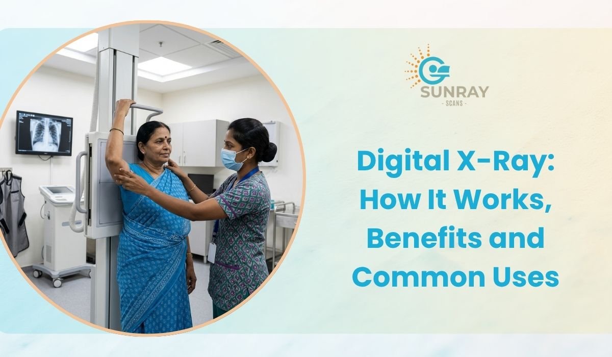
Digital X-Ray: How It Works, Benefits and Common Uses
When you slip on a wet floor, twist your ankle playing cricket, or deal with a stubborn chest cold that refuses to clear up, your doctor will almost certainly write out a prescription for an X-ray.
It is the oldest, most familiar test in medical care. For generations, having an X-ray meant standing frozen against a cold wall, waiting for a loud buzz, and sitting in a clinic hallway for an hour while a technician developed large plastic film sheets inside a chemical darkroom. Those dark, plastic sheets were then held up against a fluorescent light box on a doctor’s wall so they could search for bone cracks.
That traditional film method served us well for a century, but healthcare has moved forward. Today, traditional film has been replaced by modern digital technology.
A digital X-ray works very much like modern digital photography. Instead of relying on slow chemical film, it uses electronic sensors to capture crystal-clear pictures of your internal anatomy in a fraction of a second. The resulting images appear directly on a high-definition computer screen, giving doctors instant insights with far less radiation exposure.
If your doctor has recommended a diagnostic check and you are looking for a reliable digital X-ray in Chennai, this guide breaks down how this modern technology works, why it is so much better than old film systems, and where it is commonly used in everyday medicine.
What Is a Digital X-Ray?
An X-ray is a form of electromagnetic radiation, just like visible light, but with much higher energy. Because of this higher energy, X-ray waves can pass through soft tissues like skin, fat, and muscle, while being absorbed by denser materials like bone and metal.
A digital X-ray uses this same physical principle, but it changes how the picture is collected and processed.
In a traditional setup, the rays passed through your body and hit a sheet of photographic film coated with silver compounds. That film had to be dipped into chemical baths to make the picture visible.
In a digital setup, the chemical film is replaced by a digital detector panel. When the invisible beams pass through your body, they hit digital sensors that instantly convert the energy into electrical signals. A connected computer processes these signals within seconds, displaying a sharp, black-and-white picture of your bones, joints, and organs right on the doctor’s monitor.
How Does an X-Ray Create an Image?
Have you ever wondered why bones look bright white on an X-ray scan, while your lungs look almost completely black? It all comes down to density and absorption.
- Bones and Teeth: These structures are packed with calcium, which is very dense. Calcium absorbs a large amount of the X-ray beam, preventing the rays from reaching the digital panel behind you. Because fewer rays reach the sensor in these areas, the computer paints your bones bright white.
- Lungs and Air Cavities: Your lungs are filled with air, which has very low density. The X-ray beams pass straight through the air without stopping and hit the sensor panel with full energy. The computer paints these areas dark black.
- Soft Tissues (Muscles, Fat, Fluid): Muscles, organs, and blood vessels have a density that sits right in the middle. They absorb some rays while letting others pass through. The computer renders these tissues in varying shades of grey.
If you have pneumonia, fluid collects inside your normally air-filled lungs. On a digital image, that fluid absorbs more beams than healthy air would, showing up as cloudy grey patches that tell your doctor exactly where the infection is sitting.
6 Big Benefits of Digital X-Ray Over Traditional Film
Switching from chemical film to digital imaging has brought massive improvements for both patients and healthcare providers. Here is why modern medical clinics have completely retired older film systems:
Significantly Lower Radiation Exposure
Safety is always the primary concern whenever people hear the word radiation. One of the biggest advantages of digital technology is its exceptional sensitivity. Digital sensor panels can detect very faint beams of energy, which means the machine needs up to 70 to 80 percent less radiation to produce a crystal-clear image compared to traditional film. This makes the process much safer for children, seniors, and patients who need repeat scans.
Instant Results with Zero Waiting
With old film systems, you had to wait while the staff manually processed the plastic sheets in a darkroom with liquid chemicals. If the image came out blurry, overexposed, or too dark, the whole process had to be repeated from scratch.
With a digital scan, the image appears on the technician’s computer screen within two to three seconds after the button is pressed. The technician can instantly check that your positioning is correct, wrap up your appointment, and let you get back to your day without delays.
Superior Image Quality and Digital Enhancement
Traditional film was permanent; what was printed was what you got. If a section was slightly dark, a doctor had to squint at a light box or order another scan.
Digital images give radiologists powerful software tools. They can:
- Zoom in on tiny hairline cracks in small toe or finger bones.
- Adjust brightness and contrast to inspect both dense bone and surrounding soft tissue on the exact same image.
- Invert colors or highlight specific tissue densities for clear analysis.
- Measure bone angles and joint spaces down to the millimeter.
Eco-Friendly and Chemical-Free
Traditional film processing required toxic chemicals like fixer and developer liquids, which had to be handled carefully and disposed of to prevent environmental pollution. Digital imaging is entirely paperless and chemical-free, eliminating hazardous waste completely.
Easy Sharing and Permanent Digital Storage
Instead of carrying around large, delicate plastic envelopes that bend, tear, or fade over time, your digital files are saved securely in standard medical formats (like DICOM). Your scans can be viewed across different clinic computers, stored in your personal digital health locker, or sent directly to your specialist via email or messaging apps for a quick second opinion.
Fewer Retakes and Less Physical Discomfort
Because the software can adjust contrast and brightness digitally, technicians rarely need to take a second shot. This means less time standing in uncomfortable positions while dealing with a painful injury.
Comparing Traditional Film X-Rays with Digital X-Rays
Feature | Old Film X-Ray | Modern Digital X-Ray |
Radiation Level | Standard baseline exposure | Up to 70 to 80 percent lower exposure |
Time to View Image | 15 to 30 minutes (chemical development) | 2 to 3 seconds on a digital screen |
Image Adjustments | None (fixed on plastic film) | Zoom, contrast, brightness, and angle tools |
Storage Method | Physical plastic film sheets | Secure digital files and cloud storage |
Environmental Impact | Uses chemical developers and fixers | Completely clean and chemical-free |
Risk of Retakes | Higher due to exposure mistakes | Minimal due to digital sensor sensitivity |
Common Uses: When Do Doctors Recommend a Digital X-Ray?
A digital X-ray scan remains the go-to first-line diagnostic test across almost every field of medicine. Here are the most common situations where a doctor will request one:
Orthopedics and Bone Fractures
The most common use of an X-ray is checking bones. Whether you have had a road accident, a sports injury, or a simple misstep down a staircase, an X-ray is the fastest way to confirm:
- Bone fractures and small hairline stress cracks.
- Joint dislocations (such as a popped shoulder or finger).
- Bone alignment problems and curvature issues like scoliosis.
- Chronic joint wear, cartilage loss, and bone spurs from osteoarthritis.
Chest and Respiratory Health
A chest X-ray is one of the most widely performed diagnostic tests in the world. It provides a quick look at your heart, lungs, and chest wall. Doctors use it to diagnose:
- Pneumonia and chest infections (visible as fluid buildup).
- Tuberculosis, showing distinct patterns of tissue changes.
- Chronic Obstructive Pulmonary Disease (COPD) and emphysema.
- Fluid around the lungs (pleural effusion).
- Enlarged heart size (cardiomegaly), which can indicate underlying heart strain.
Dental Care
Dentists rely heavily on digital dental X-rays to check the health of your teeth beneath the gumline. These scans reveal hidden cavities between teeth, check for infections at the tooth root, monitor wisdom teeth growth, and guide dental implant planning.
Abdominal Discomfort and Digestive Issues
If a patient arrives with severe belly pain, persistent vomiting, or bloating, an abdominal X-ray helps doctors spot:
- Intestinal blockages or twisted bowel loops.
- Perforations in the stomach or intestines, visible as trapped air pockets under the diaphragm.
- Kidney stones or gallstones that contain enough calcium to show up clearly.
Detecting Swallowed Objects
Toddlers and young children frequently swallow small foreign items like coins, small toy parts, button batteries, or safety pins. A quick, low-dose digital scan tracks down the exact location of the item in the throat, stomach, or digestive tract so doctors can decide whether it will pass safely or needs removal.
Pre-Operative and Employment Clearances
Surgeons frequently order a routine chest X-ray before major operations under general anesthesia to ensure the patient’s heart and lungs are strong and healthy. Similarly, routine chest imaging is often required for visa applications and company health checks to screen for infectious respiratory conditions.
What to Expect During Your X-Ray Appointment
One reason the X-ray has remained the primary medical test for over a century is that it is fast, simple, and completely painless.
Simple Preparation Steps
- Wear Loose Clothing: Choose comfortable, loose cotton clothing. Avoid clothes with metallic embroidery, metal zippers, hooks, or shiny buttons, as metal stops X-rays and leaves white spots that can hide underlying bone details.
- Remove Metal Accessories: Take off watches, jewelry, eyeglasses, belts with metal buckles, hairpins, and body piercings around the area being scanned.
- Inform About Pregnancy: Always inform the technician if you are pregnant or think you might be. While the radiation from a modern digital machine is minimal, extra care is taken to protect a developing baby, usually by using a protective lead apron or choosing an alternative test like ultrasound.
The Scanning Procedure
- Positioning: The technician will guide you to stand against a sensor board, sit on a chair, or lie down on a cushioned table depending on which body part needs imaging.
- Lead Shielding: The staff may place a heavy lead apron over your lower body to shield reproductive organs from unnecessary scattered rays.
- Staying Still: The technician will step behind a protective glass window and ask you to hold completely still for just two seconds. For a chest image, they will instruct you to take a deep breath in and hold it.
- Completion: The machine emits a quiet click, and the scan is done.
The entire process takes less than five to ten minutes from the time you walk into the room until you walk out.
A Quick Note on Radiation Safety
We are exposed to natural background radiation every day from the sun, the soil, and building materials. A standard digital chest X-ray exposes you to about the same tiny amount of radiation you naturally absorb from the environment over just two to three days. The health risks of missing a serious bone break or lung infection are far higher than the tiny, safe dose used in modern digital imaging.
How to Choose the Right X-Ray Centre in Chennai
While X-rays are widely available across clinics, hospitals, and diagnostic centers, the quality of the technology and the expertise of the staff make a real difference in the care you receive.
When looking for a digital X-ray in Chennai, look for an X-ray centre Chennai residents trust for:
- Modern Digital Equipment: Centers using high-frequency digital radiography units that offer minimal radiation doses and high image clarity.
- Certified Radiographers: Skilled staff who position you gently and accurately the very first time, avoiding painful repositioning.
- Fast Digital Reports: Clinics that provide instant digital copies, high-definition prints, and rapid reporting so your doctor can start treatment immediately.
- Clean and Hygienic Facilities: Sanitized equipment, comfortable waiting spaces, and clear patient guidance.
Why Choose Sunray Scans for Your X-Ray in Chennai
When you need an accurate diagnosis for yourself or a family member, you want a medical center that blends technical accuracy with a gentle, patient-focused approach.
For reliable diagnostic care, visit Sunray Scans, a leading medical imaging and diagnostic center located in Royapettah, Chennai.
Sunray Scans features advanced, low-dose digital radiography systems alongside a complete range of diagnostic services, including high-resolution Open MRI, multi-slice CT angiography, computerized ultrasound, and automated laboratory tests. Their experienced team of radiographers and senior radiologists ensures that every scan is captured with precision, providing clear reports that your doctor can rely on with complete confidence.
What Sets Sunray Scans Apart?
- Low-Dose Digital Radiography: Cutting-edge digital sensor panels that deliver sharp images with reduced radiation.
- Quick and Walk-In Friendly: Fast walk-in services with minimal waiting times, making urgent checks quick and stress-free.
- Experienced Radiologists: Comprehensive evaluations and quick turnaround times so you can consult your treating physician without delay.
- Comprehensive Health Services: A full suite of companion checks, including blood tests, ECG, and advanced scans, all available under one roof.
- Transparent and Affordable Rates: High-quality healthcare delivered at fair, accessible prices for families across Chennai.
A simple X-ray is often the very first step toward healing and peace of mind. Whether you are dealing with a sports injury, chronic joint stiffness, or a stubborn cough, getting a clear picture of what is happening inside helps you get back to feeling your best. Talk to your doctor, choose a reliable diagnostic partner, and take care of your health today.